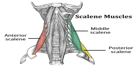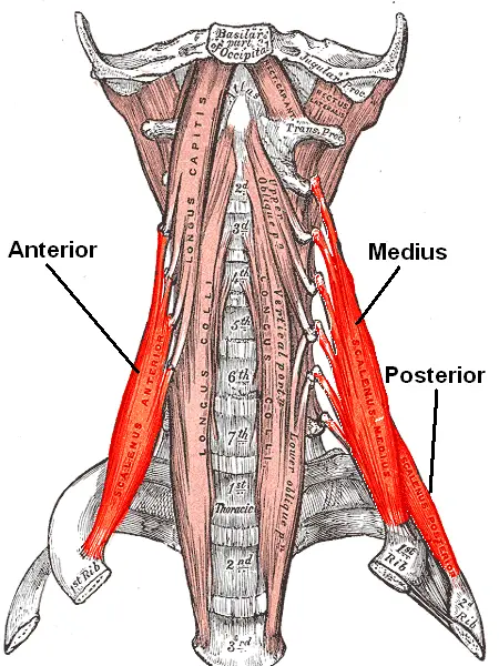
Scalene Anterior Muscle
Scalene anterior Muscle is a group of three pairs of muscles in the lateral neck, namely the anterior scalene. It is located deeply just behind the Sternocleidomastoid muscle. The brachial plexus and subclavian artery runs between the anterior and middle scalene muscles. This provides an important anatomical locations for anaesthetics to do an interscalene block….