Arytenoid Muscle
Table of Contents
Arytenoid Muscle Anatomy
Arytenoid muscle is a single muscle, that fills up the posterior concave surfaces of the arytenoid cartilages.
The arytenoid muscle, also known as the interarytenoid muscle, is a composite intrinsic muscle of the larynx that is made up of two parts: an unpaired transverse arytenoid muscle and a bilaterally paired oblique arytenoid muscle. The two portions can be thought of as independent muscles.
The attachments, structures, and actions of the two-component pieces are different. The recurrent laryngeal nerve(s), which are branches of a single vagus nerve (CN X), innervate both of them motorly.
Origin:
Arytenoid cartilage on one side.
Insertion:
Arytenoid cartilage on opposite side.
Nerve:
recurrent laryngeal branch of the vagus.
Actions
approximate the arytenoid cartilages (close rima glottis).
Relations
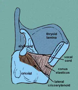
The sole unpaired intrinsic muscle of the larynx is the transverse arytenoid. It is situated directly above the cricoid cartilage on the posterior part of the larynx. It fills the concavities on the posterior surface of the arytenoid cartilages and closes the space between them. The two oblique arytenoid muscles are located superficially to the transverse arytenoid muscle. When combined, the three muscles are referred to as the arytenoid, or interarytenoid, muscle. The transverse and oblique arytenoids are sometimes thought of as two segments of the same muscle.
The circular (constrictor) and longitudinal muscles of the pharynx are located externally to the transverse arytenoid and other intrinsic laryngeal muscles. The pharynx and larynx are encircled by a semicircular tube made up of these muscles.
Clinical Significance
Electromyography
An effective way to assess recurrent laryngeal nerve function is through examination of the arytenoid muscle’s activity. Surgeons can have peace of mind knowing that during neck surgeries like thyroidectomies, the recurrent laryngeal nerve is not harmed by continuous electromyography of the arytenoid muscle.
Dogs are among the many species that have the arytenoid muscle.
FAQ
The larynx’s intrinsic, unpaired transverse arytenoid muscle. It covers the posterior side of the arytenoid cartilage and is located just superior to the cricoid cartilage. Being an adductor of the vocal fold, the transverse arytenoid is crucial to phonation.
the larynx
The larynx contains paired, pyramid-shaped cartilage structures called arytenoid cartilages, which are crucial for producing voice tone. They aid in the formation of the cricoarytenoid joints and are situated on the lateral portion of the superior border of the cricoid cartilage lamina.
The arytenoid cartilages are adducted by the transverse and oblique arytenoids muscles, which closes the posterior region of the rima glottidis. The laryngeal inlet narrows as a result.