
Brachioradialis muscle
Table of Contents
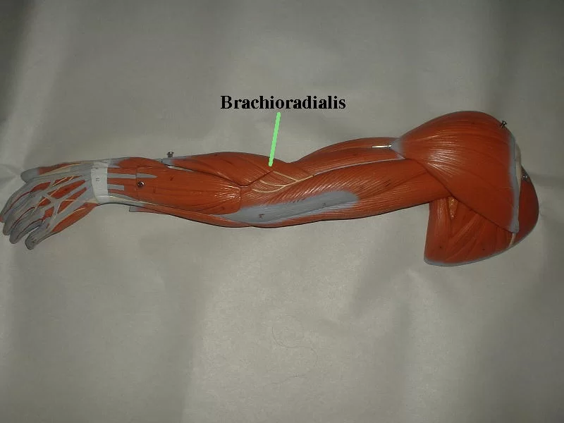
Brachioradialis Muscle Anatomy
The brachioradialis is a muscle of the forearm that flexes the forearm at the elbow. It is also capable of both pronation and supination, depending on the position of the forearm.
Origin
It originates from the lateral supracondylar ridge of the humerus.
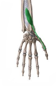
Insertion
It inserts into the distal radius (radial styloid process).
Nerve supply
The radial nerve supplies the muscle.
Blood Supply
The radial artery supplies the muscle.
Actions
It flexes the elbow and supinates and pronates the radioulnar joint at 90′.
Function
The brachioradialis muscle works in synergy with the biceps brachii and brachialis to flex the forearm at the elbow. Brachioradialis is a powerful forearm flexor when the forearm is semi-pronated, meaning that the palm is perpendicular to the ground.
When the forearm is positioned halfway between supination and pronation at the radioulnar joint, the brachioradialis is a more powerful elbow flexor. Because the biceps brachii are mechanically disadvantaged when the elbow flexes when pronated, the brachioradialis is more active.
The brachioradialis does not produce as much joint torque as the brachialis or the biceps because of its location so far from the elbow’s fulcrum. It works best when the muscles at the elbow have already partially flexed. while rapid movement is needed or while lifting a weight during gradual forearm flexion, the brachioradialis flexes the forearm at the elbow.
When in a midposition, such as when hammering, the muscle is required to stabilize the elbow during rapid flexion and extension. While the triceps brachii and anconeus are antagonistic, the brachioradialis works in concert with the brachialis and biceps brachii.
Relations
The muscle closest to the skin on the radial aspect of the forearm is the brachioradialis. When the forearm is flexed and semi-pronated, it is easily palpable as the fleshy protrusion in the upper part of the lateral forearm. The muscle defines the boundary between the anterior and posterior forearm compartments and forms the lateral wall of the cubital fossa.
The tendon of the biceps brachii goes deep to its proximal section on its route to the radial tuberosity, whereas the distal part of the brachialis muscle covers its medial side. Between the brachioradialis and brachialis travel the radial nerve and the vascular anastomosis between the radial recurrent and deep brachial (profunda brachii) arteries.
The superficial surface of the muscle is crossed by the lateral cutaneous antebrachial nerve and the cephalic vein. The flexor carpi radialis muscle is lateral to it in the mid-forearm.
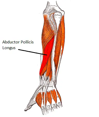
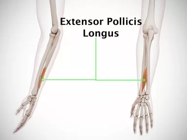
The tendon is lateral to the radial artery at the wrist level. Take note that the radial pulse may be felt at this location. The radial nerve’s superficial branch also emerges deep to the brachioradialis at this location. Before superficially passing over the extensor retinaculum to enter the hand, it travels between this and the extensor carpi radialis brevis muscle. The tendons of the extensor pollicis brevis and abductor pollicis longus muscles cross the brachioradialis tendon just proximal to its insertion.
Assessment
The Brachioradialis muscle was Palpated on the anterolateral surface of the forearm.
In order to focus on the brachioradialis, we will ask the patient to bend their forearm in a mid-position, which involves bending their elbow against resistance. This is how the MMT is typically performed for the three main elbow flexors: the biceps, brachialis, and brachioradialis.
It should be noted that during the test, the wrist flexor muscles should stay relaxed because tight wrist flexor contractions may aid in elbow flexion.
Strengthening Exercises
1. Resisted band supination
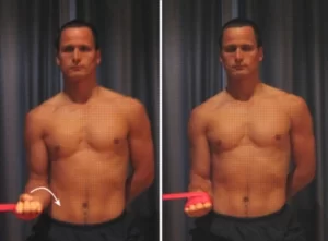
- Begin this exercise with a resistance band around the hand.
- The elbow should be rested at the side and bent to 90 degrees.
- Slowly rotate the forearm against the resistance band so the palm faces up.
- Perform 3 sets of 10 repetitions as far as possible and comfortable without pain.
2. Resisted band pronation
- Begin this exercise with a resistance band around the hand.
- The elbow should be rested at the side and bent to 90 degrees.
- Slowly rotate the forearm against the resistance band so the palm faces down.
- Perform 3 sets of 10 repetitions as far as possible and comfortable without pain.
Stretching Exercises

- Straighten the right arm and form a fist. Grasp the right fist in the left hand.
- Rotate the right arm until the fisted thumb points towards the body.
- Keeping the arm straight use the left hand to gently flex the right wrist. This is the same motion one would make during wrist curls, bringing the palm closer toward the forearm.
- Hold the stretch for 15 to 30 seconds, breathing normally, then repeat on the other side.
Related pathology:
If this muscle is too tight and/or contains trigger points, it can send pain to your elbow and forearm. Additionally, you might feel pain on the back of your hand or between your thumb and index finger. Thus it may contribute to the tennis elbow pain.
Brachioradialis pain is usually a shooting pain in your forearm or elbow. It’s often confused with tennis elbow. While both are typically caused by overuse and overexertion, tennis elbow is an inflammation of the tendons in your elbow, and brachioradialis pain is specific to this muscle.
When the brachioradialis and extensor carpi radialis longus tendon compress the superficial radial nerve, a condition known as Wartenberg syndrome develops. Forearm pronation causes more compression on the radial nerve. Only burning pain and paresthesia over the dorsum of the wrist, hand, and dorsal surface of the thumb, index, and middle fingers are the sensory manifestations that arise from it.
The C6 spinal nerve root is tested clinically using the brachioradialis tendon. which is impacted by disc herniation in the C5–C6.
The brachioradialis and extensor carpi radialis longus are the first two muscles to regain innervation when the radial nerve is injured in the radial groove, which makes them crucial to assess for radial nerve recovery in cases of midshaft fracture of the humerus.
6 Comments