Lateral Cricoarytenoid Muscle
Table of Contents
Lateral cricoarytenoid muscle Anatomy
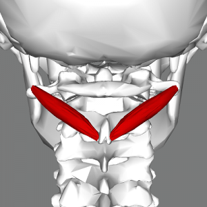
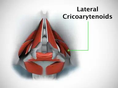
The lateral cricoarytenoid muscles extend from the lateral cricoid cartilage to the muscular process of the arytenoid cartilage. the arytenoid cartilages medially, these muscles adduct the vocal cords and close the rima glottidis protecting the airway.
Muscle details:
Extend from the lateral cricoid cartilage to the muscular process of the arytenoid cartilage.
Sound generation is facilitated by the larynx’s intrinsic muscles. They accomplish this by adjusting the larynx’s numerous components, altering the vocal folds’ length and tension, and opening or shutting the rima glottidis. More specifically, by adducting the vocal folds, the lateral cricoarytenoid muscle helps produce sound.
Origin:
the lateral part of the arch of the cricoid.
Insertion:
Muscular process of the arytenoid cartilage.
Nerve supply:
recurrent laryngeal branch of the Vagus Nerve.
Blood Supply
The superior and inferior thyroid arteries’ laryngeal branches supply blood to this muscle.
Action:
Adduct and medially rotate the cartilage, pulling the vocal ligaments towards the midline and backward and so closing off the rima glottidis.
Function
The lateral cricoarytenoid is a member of the intrinsic laryngeal muscles, which are all involved in phonation, or the creation of sound. They accomplish so by manipulating the various sections of larynx; changing the state of the vocal folds (i.e. tension and length) and opening or shutting the rima glottidis.
The vocal process, to which the vocal ligaments are linked, is medially swung when the lateral cricoarytenoid muscles contract, rotating the arytenoid cartilages. As a result, the vocal folds are brought together, closing the anterior portion of the rima glottidis, and the tips of the vocal processes come together.
Applying this to speech: the transverse and oblique arytenoid muscles simultaneously pull the arytenoid cartilages toward one another, sealing the posterior region of the rima glottidis; this is known as adductive tension, while the lateral cricoarytenoid rotates the arytenoid cartilages. The laryngeal intake narrows, the rima glottidis shuts, and the vocal folds adduct collectively. Vocal folds that have been adducted vibrate and make voiced sounds when expired air travels through the closed rima glottidis and reaches the folds.
The lateral cricoarytenoid cannot completely shut the rima glottidis when acting alone. A portion of the air goes through the vocal folds without fully resonating them. Whispering results from this.
Relations
The vocal folds are lateral to the muscle. Since they all insert into the muscular process of the arytenoid cartilage, its insertion is closely related to that of the transverse arytenoid and posterior cricoarytenoid muscles.
Clinical Importance
Laryngeal Hemiplegia
An uncommon but common condition known as laryngeal hemiplegia (paralysis), sometimes known as “roaring in horses,” is defined by atrophy of the dorsal and lateral cricoarytenoid muscles (abductor and adductor of the arytenoid cartilage), especially on the left side. Primary denervation (recurrent laryngeal neuropathy) of no known cause (idiopathic axonopathy) and, to a much lesser extent, subsequent nerve injury are the most common causes of muscular atrophy.
The abnormal inspiratory sounds (roaring) during exercise in horses with laryngeal hemiplegia are caused by paralysis of the left dorsal and lateral cricoarytenoid muscles, which induce incomplete dilatation of the larynx, restriction of airflow, and vibration of vocal cords.
FAQ
During deglutition, vocalization, and expiration, the arytenoid on the cricoid cartilage can rotate inward thanks to the lateral cricoarytenoid muscle, which closes the laryngeal airway. The arytenoids are drawn together by the interarytenoid muscle, which increases this activity.
In order to produce sound, the LCA muscle moves the vocal cords from an open-breathing position to a closed position. Within the vocal cord, this muscle is situated directly lateral to and parallel to the TA muscle.
Only the posterior cricoarytenoid muscles are used for abduction. They open the glottis by drawing the posterior ends of the arytenoid cartilages together. The vocal folds are pulled apart as a result of this pulling apart the front ends, which is where they attach. These adductors are called lateral cricoarytenoid.
the larynx
An intrinsic muscle of the larynx is the lateral cricoarytenoid (also known as the anterior cricoarytenoid). It connects anteriorly at the cricoid cartilage and posteriorly at the arytenoid cartilage on the same side. It functions to block the airway by closing the rima glottidis.
The vocal folds become tight from its isometric contraction, which improves regular vibration. The interarytenoid and lateral cricoarytenoid muscles are the traditional adductor muscles.