
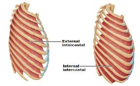
External intercostal muscles
External intercostal muscles are eleven in number on both sides.
origin:
Lower border of ribs.
Insertion :
Upper border of rib below.
Nerve:
intercostal nerves.
Actions:
Inhalation.
External intercostal muscles are eleven in number on both sides.
origin:
Lower border of ribs.
Insertion :
Upper border of rib below.
Nerve:
intercostal nerves.
Actions:
Inhalation.
Physiotherapist , Samarpan Physiotherapy Clinic, Vastral, Nirant Cross Road, Ahmedabad
Home Visit Treatment Also Available in Bapunagar Vastral Rabari Colony Char Rasta, CTM, Maninagar , Viratnagar , Nikol Nava Naroda And NearBy Area Of Ahmedabad.
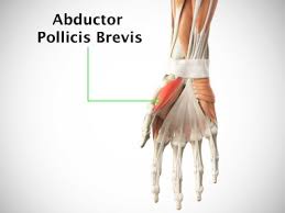
Abductor Pollicis Brevis Muscle Anatomy The abductor pollicis brevis is a flat, thin muscle located just under the skin. It is a thenar muscle and therefore contributes to the bulk of the palm’s thenar eminence. Origin It originates from the flexor retinaculum of the hand, the tubercle of the scaphoid bone, and additionally sometimes from…
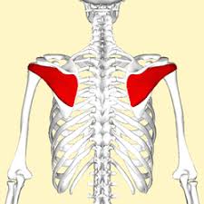
Muscle details : The infraspinatus muscle is a thick triangular muscle, which occupies the chief part of the infraspinatous fossa. It is one of the muscles of rotator cuff. Origin : It originates from the medial two -thirds of the infraspinous fossa of the scapula . Insertion : It inserts into the middle impression on…
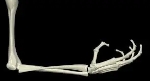
Introduction A human arm is composed of three bones, namely – the humerus, ulna, and radius. The human arm is an essential component that allows movement along the shoulder, elbow, wrist, and fingers, which is helpful for daily tasks. The humerus, ulna, and radius are the three bones that make up an arm in humans….
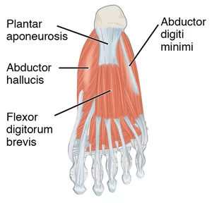
Abductor hallucis muscle is an intrinsic muscle of the foot. It works mainly in the abduction and flexion of the great toe. origin: Medial process of calcaneal tuberosity, Plantar aponeurosis, Flexor retinaculum . Insertion: Medial aspect of the base of 1st phalanx of the hallux. Nerve: Medial plantar nerve. Blood supply: Medial plantar and first…
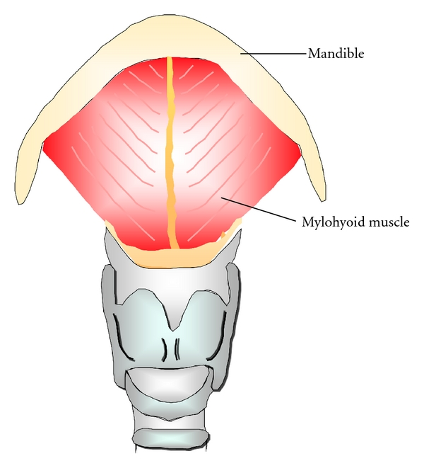
Introduction The mylohyoid muscle is one of the suprahyoid muscles.The mylohyoid muscle is a flat, triangular muscle lying deep in the anterior belly of the digastric. Both the sides of the mylohyoid muscles together form the floor of the mouth. Along with the other suprahyoid muscles (the digastric, the geniohyoid, and the stylohyoid), The mylohyoid…
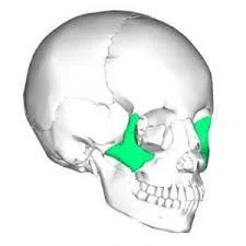
Introduction The zygomatic bones are also referred to as the cheekbones. These bones are located directly beneath each eye and extend upward to the outer side of each eye. The zygomatic bones connect to several other facial bones, including the nose, jaw, portions of the eye, and bones just in front of the ears. The…