Iliococcygeus Muscle
Iliococcygeus Muscle Anatomy
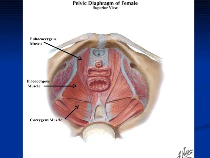
The levator ani muscle, which makes up the majority of the pelvic diaphragm, is composed of three muscles, including the iliococcygeus muscle. The flat, broad skeletal muscle is known as the iliococcygeus.
the inner side of the ischium (the lower and back part of the hip bone) and from the posterior part of the tendinous arch of the obturator fascia, and is attached to the coccyx and anococcygeal body; it is usually thin and may be absent, or be largely replaced by fibrous tissue. An accessory slip at its posterior part is sometimes named the iliosacralis.
Three muscles make up the levator ani muscle group:
- Puborectalis (puboanalis),
- Pubococcygeus
- Iliococcygeus.
Origin:
ischial spine and from the posterior part of the tendinous arch of the pelvic fascia.
After meeting the fibers from the opposing side, the iliococcygeus creates the midline raphe, which is continuous with the anococcygeal ligament and offers a stable anchoring point for the pelvic floor.
Insertion
coccyx and anococcygeal raphe.
Nerve supply:
levator ani nerve (S4).
inferior rectal nerve from pudendal.
Blood Supply
The branches of the inferior gluteal, inferior vesical, and pudendal arteries give blood to the iliococcygeus muscle.
Action
supports the viscera in the pelvic cavity.
The iliococcygeus muscle, which is a component of the pelvic diaphragm, raises the pelvic floor and offers structural support to nearby pelvic structures. When at relaxation, its fibers can retain a tonic contraction, which relaxes throughout the defecation process.
Clinical Importance
- Pelvic Organ Prolapse
- Urinary incontinence
- Bowel incontinence