
Knee Joint: Anatomy, Function, Importance, Exercise
Table of Contents
Introduction:
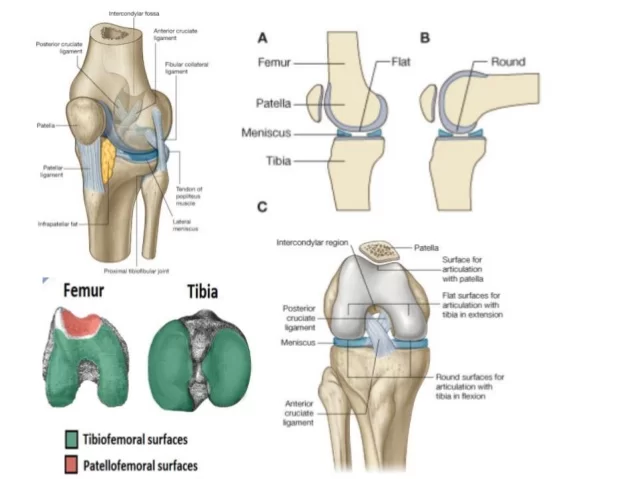
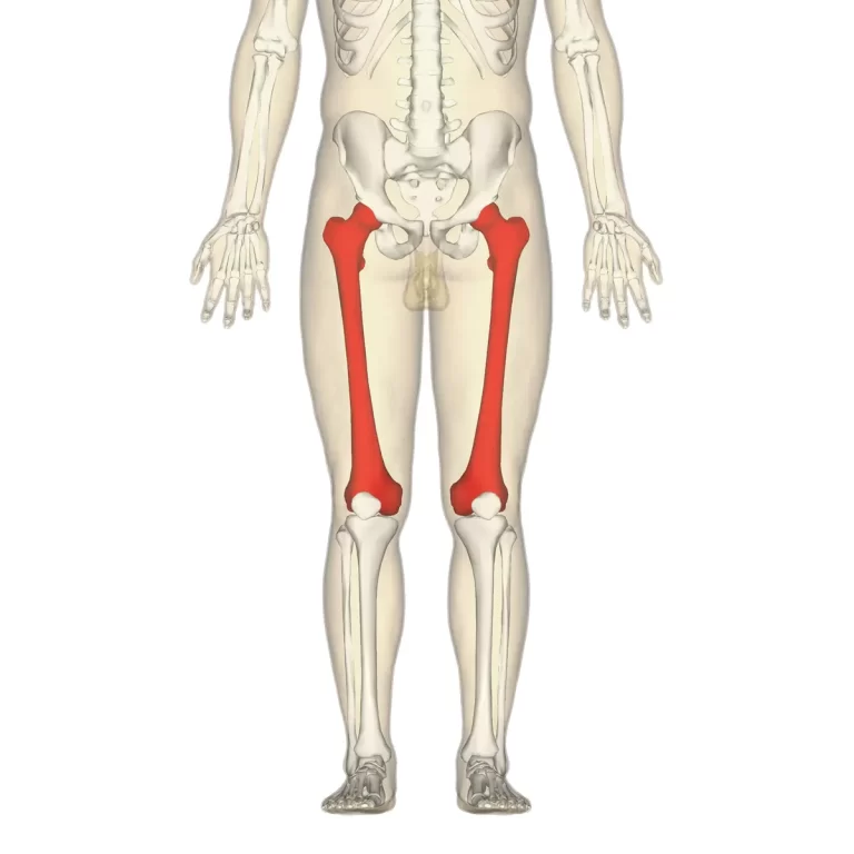
knee joint is the largest and most complex joint of the body. The knee is one of the largest and most complex joints in the body. The knee joins the femur to the tibia. The smaller bone that runs alongside the tibia and the patella are the other bones that make the knee joint.
Type of Knee Joint:
~It is a compound synovial joint.
Joints:
~it consists of 3 joints.
1. Medial condylar joint:-between the medial condyle of the femur and medial condyle of the tibia.
2. Lateral condylar joint:-between the lateral condyle of the femur and lateral condyle of the tibia.
3. Patellofemoral joint:-between the patella and patellar surface of the femur.
Articular surface:
- The condyles of the femur.
2. The patella.
3. The condyles of the tibia.
Femoral condyles:
*.Lateral condyle:
~small radius of curvature
~smaller in all dimensions
~extends more anteriorly
*Medial condyle:
~larger radius of curvature
~extends more distally
*intercondylar notch
*Trochlear groove and intercondylar notch:
~Anteriorly, the condyles are separated by a patellofemoral groove.
~posteriorly, the condyles are separated by the intercondylar notch.
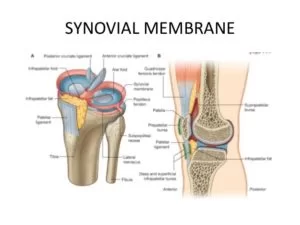
~The synovial membrane of the knee joint attaches to the margins of the articular surfaces and to the superior and inferior margins of the menisci.
~it lines the joint capsule except posteriorly where the cruciate ligament is found.
~in front, it is absent from the patella.
~the two cruciate ligaments, which attach in the intercondylar region of the tibia below and the intercondylar fossa of the femur above are outside the articular cavity but inclosed within the fibrous membrane of the knee joint.
~posteriorly, the synovial membrane reflects off the fibrous membrane of the joint capsule on either side of the posterior cruciate ligament and loops forward around both ligaments thereby excluding them from the articular cavity.
~Anteriorly, the synovial membrane is separated from the patellar ligament by an infrapatellar fat pad.
~alar fold
~the infrapatellar synovial fold
~pouches in two locations
~subpopliteal recess
~suprapatellar bursae
Bursae of Knee Joint:
~13 bursae have been described around the knee joint
~the four are anterior
~four are lateral
~five are medial
*Anterior bursae:
~subcutaneous prepatellar bursae
~subcutaneous prepatellar bursae
~deep infrapatellar bursae
~suprapatellar bursae
*Lateral bursae:
~a bursae deep to the lateral head of the gastrocnemius.
~a bursae b/w fibular collateral ligament and the biceps femoris.
~ bursae b/w fibular collateral ligament and tendon of the popliteus.
~ a bursa b/w tendon of popliteus and lateral condyle of the tibia.

*Medial bursae:
~a bursae deep to the medial head of the gastrocnemius.
~the anserine bursae
~a bursae deep to the tibial collateral ligament
~a bursae deep to semimembranosus.
~ocassionally fifth bursae present b/w tendons of semimembranosus and semitendinosus.
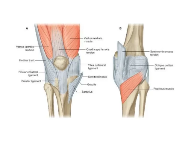
Ligaments of Knee joint:
~fibrous capsule
~ligamentum patellae
~tibial collateral or medial ligament
~fibular collateral or lateral ligament
~oblique popliteal ligament
~arcuate popliteal ligament
~anterior cruciate ligament
~posterior cruciate ligament
~medial meniscus
~lateral meniscus
~transverse ligament
Fibrous capsule:
~thin capsule with tibial and femoral attachment
~anteriorly deficient
~popliteus muscle and tendon
*Fibrous capsule strengthening:
~anteriorly-medial and lateral patellar retinacula(vastus medialis ,vastus lateralis)
~laterally:-illiotibial tract
~medially:-tendons of sartorius,semimembranosus
~posteriorly:-oblique popliteal ligament
*Coronary ligament:-
~fibrous capsule is attached to the periphery of the menisci.
~connects the periphery of the menisci to the tibia.
~they are a portion of the capsule that is stressed in rotary movements of the knee.
*fibrous capsule opening:-
~two constant gaps
~leading into suprapatellar bursae
~exit of popliteal tendon
~sometimes there are gaps that communicate with bursae deep to the medial head of the gastrocnemius and deep to the semimembranosus.
*Ligamentum patellae:-
~it is the central portion of the common tendon of insertion of quadriceps femoris(remaining portions of the tendon from medial & lateral patellar retinacula).
~it is related to superficial and deep infrapatellar bursae and infrapatellar fat of the pad.
~attachments:
~above:-apex of patella
~below:-tibial tuberosity
*Cruciate ligament:
~very thick, strong fibrous bands.
~direct bonds of union between femur and tibia.
~represent collateral ligament of original femorotibial joints.
~maintain anteroposterior stability.
~named according to the attachment on the tibia.
~supplied by vessels and nerves that pierce oblique popliteal ligament.
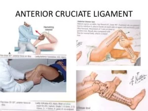
*Anterior cruciate ligament:
~the anterior cruciate ligament attaches to a facet on the anterior part of the intercondylar area of the tibia and ascends posteriorly to attach to a facet at the back of the lateral wall of the intercondylar fossa of the femur.
~the anterior cruciate ligament crosses laterally to the posterior cruciate ligament as it passes through the intercondylar region.
~the anterior cruciate ligament prevents anterior displacement of the tibia relative to the femur.
~it is taut during knee extension.
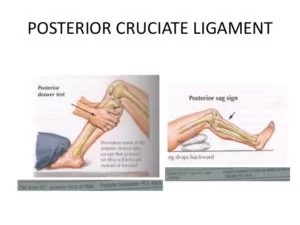
*Posterior cruciate ligament:
~the posterior cruciate ligament attaches to the posterior aspect of the intercondylar area of the tibia and ascends anteriorly to the medial wall of the intercondylar fossa of the femur.
~posterior cruciate ligament restricts posterior displacement.
~it taunts during knee flexion.
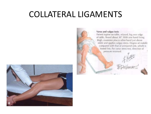
*Medial collateral ligament:
~is attached superiorly to the medial epicondyle of the femur just below the adductor tubercle.
~inferiorly it divides into superficial and deep.
~superficial part attached to the upper third of the tibia, as far down as the tibial tuberosity.
~the deep portion, which is short, fuses with the capsule and with the medial meniscus.
~A bursae usually separates the two parts.
~MCL, tightens in extension.
~a valgus stress will put a strain on the ligament.
*Lateral collateral ligament:
~superiorly attached to the lateral condyle of the femur just above the popliteal groove.
~inferiorly embraced with the tendon of the biceps femoris and attached to the head of the fibula in front of its apex.
~separated from lateral meniscus by popliteal tendon and fibrous capsule.
~infeolateral genicular vessels and nerves separate it from the capsule.
~tightest in extension,0-30 degrees.
~becomes looser in flexion >30 degrees.
~primary restraint to varus.
~secondary restraint to ER and posterior translation.
*Oblique popliteal ligament:
~it is an expansion from the semimembranosus tendon close to its insertion into the tibia.
~oblique popliteal ligament passes upwards and laterally.
~fuses with the fabella if present.
~lends with the posterior surface of the capsule above the lateral femoral condyle.
~pierced by middle genicular vessels and nerve.
~branch from the posterior division of the obturator nerve pierces the ligament, supplies cruciate and articular twig to the knee(referred pain from pelvic peritoneum to knee)
~popliteal artery lies on it.
~strengthens the posterior portion of the capsule and prevents extreme lateral rotation.
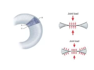
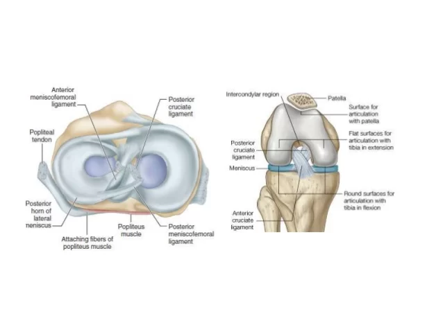
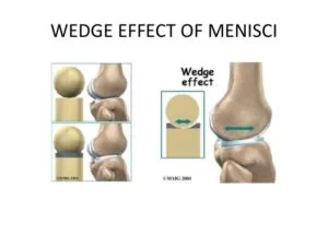
Anatomy of menisci:
~menisci are fibro cartilaginous.
~ crescent-shaped attached ends to the tibia, deepen the articular surface of the tibia.
~wedge shaped on cross-section.
~outer border thick, convex, fixed, and vascular.
~inner border thin, concave, free, avascular, and nourished by synovial fluid.
~the major orientation of collagen fibers in the meniscus is circumferential, radial fibers and perforating fibers also are present.
~the circumferential tension in the menisci counteracts this outward or radial force.
~these hoop forces are transmitted to the tibia through the strong anterior and posterior attachments of the menisci.
~hoop tension is lost when a single radial cut or tear extends to the capsular margin, in terms of load bearing, a single radial cut through the meniscus may be equivalent to meniscetomy.
*Anatomy of menisci:
~it has two ends, two borders, and two surfaces.
~flexion and extension take place at the upper surface of the menisci.
~rotation occurs between the lower surface of the menisci and the tibia.
*Medial meniscus:
~it is relatively immobile.
~it is a C-shaped, semicircular, fibrocartilagenous disc.
~peripheral margin adherent to the tibial collateral ligament.
~more liable to injury.
*Lateral meniscus:
~it is more round/circular in shape.
~the posterior end of the meniscus is attached to the femur through 2 meniscofemoral ligaments.
~the tendon of the popliteus and fibrous capsule separates it from LCL.
~Mobility of the posterior end is controlled by the popliteus and 2 meniscofemoral ligaments.
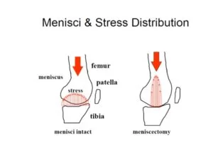
*Functions of menisci:
~shock absorption
~redistributes forces
~spread synovial fluid
~minimal effect on stability
~on rotation menisci move with the femur.
~lateral moves 20-24 mm.
~medial less mobile 10-15 mm.
~lateral meniscus bears more load.
*Transverse ligament:-
~it connects the anterior ends of the medial and lateral menisci.
*meniscofemoral ligament:-
~the anterior meniscofemoral ligament is attached to the medial aspect of the medial femoral condyle in front of PCL.
~the posterior meniscofemoral ligament is attached posterior to PCL.
~the posterior meniscofemoral ligament is usually present.
~vary in size.
*Short lateral ligament:
~extends from the lateral epicondyle of the femur.
~to the medial border of the apex of the fibula.
~it is a cord-like thickening of capsule deep to LCL.
~deep in the interval between the iliotibial band and biceps femoris.
~surrounded by biceps femoris.
*Arcuate ligament:
~its posterior expansion of the short lateral ligament.
~ it extends backward from the head of the fibula, arches over the popliteal tendon, and is attached to the posterior border of the intercondylar area of the tibia.
~fibers oriented in various directions.
~Y-shaped configuration over popliteus.
~medial limb terminates into oblique popliteal ligament.
~lateral limb, invariable present, and is less distinct.
*Fabella:-
~fabella lies at the point on the poster lateral side of the knee.
~where multidirectional collagenous tensile stress meets.
~8%-10% osseous
~90-92% cartilaginous
*poster lateral corner:
~posterior horn of the lateral meniscus.
~arcuate complex.
~popliteus
~lateral head of gastrocnemius
*Relations of knee:
*Anteriorly:
~anterior bursae
~ligamentum patellae
~patellar plexus.
*posteriorly:
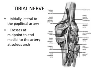
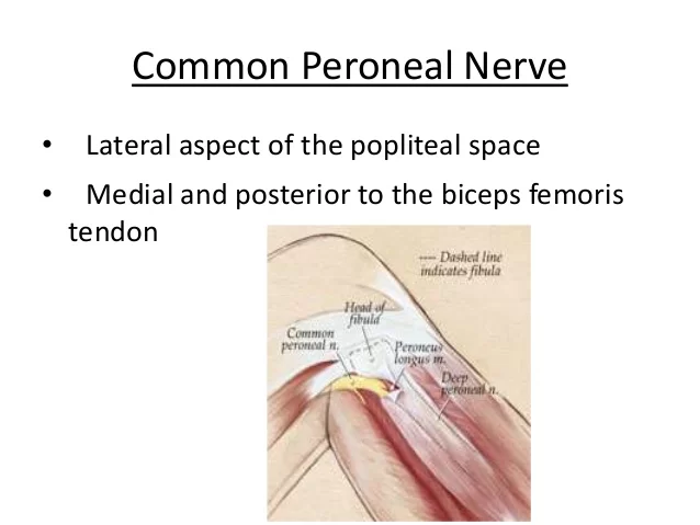
~poplitealvessel, tibial nerve, peroneal nerve, gastrocnemius, plantaris, semitendinosus, semimembranosus, gracilis, popliteus
*Popliteal fossa:
*Borders:
~superomedial:-semimembranosus
~superolateral:-biceps femoris
~inferomedial:-medial gastro head
~inferolateral:-lateral gastro head
*contents:
~popliteal artery and vein
~tibial and common peroneal nerves
*Medially:
~sartorius,semimembranosus,gracilis,saphenous vein,saphenous nerve,semimembranosus
*Laterally:
~biceps femoris
~tendon of popliteus
*Iliotibial tract:
~the iliotibial tract is a thickening of the deep fascia of the thigh, tensor fasciae lata(inserted into the tract)
~the superficial three-quarters of the gluteus maximus end in a thick tendinous lamina which is inserted into the iliotibial tract.
~it bands continuously distally to from the:-
*It tract:
~inserts distally on the greater tubercle and on a distal femur through the intermuscular septum.
~the tract is attached to the greater tubercle on the anterolateral tubercle on the anterolateral aspect of the lateral tibial condyle.
~the iliotibial band acts as an extensor of the knee when the knee is flexed from 0 to 30 degrees and as a flexor when the knee is flexed more than 40 degrees, due to the change in the transverse axis which occurs at 30-40 degree flexion.
~the pelvic tilt is a mechanism for tightening the iliotibial band. the pull of the band stabilizes the knee in extension, as well as helps to resist extension and adduction of the hip of the weight-bearing leg.
*it band biomechanics:
*Functions:
~stabilizes against varus opening.
~knee extensor in extension.
~knee flexor in flexion.
~external roatator of the tibia in >40 flexion.
*iliopatellar band:
~inserts on lateral patella resisting medially directed forces.
*Blood supply of knee:
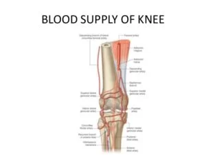
~ The knee joint is supplied by anastomoses around it.
~5 genicular branches of the popliteal artery.
~descending genicular branch of the femoral artery.
~descending branch of the lateral circumflex femoral artery.
~2 branches of the anterior tibial artery.
~circumflex fibular branch of the tibial artery.
*Lymphatic drainage of the knee:
~Drainage is to popliteal lymph nodes.
~usually 6 small nodes.
~termination of the short saphenous vein.
~popliteal artery and posterior knee (direct vessels from knee joint)
~accompanying genicular arteries(most vessels)
Nerve supply:-
~following nerves supply the knee joint.
~femoral nerve through its branches to vasti(esp vastus medialis).
~sciatic nerve through genicular branches of the tibial and common peroneal nerve.
~obturator nerve through its posterior division.
~infrapatellar branch of saphenous.
*Arterial supply of knee:-
*Femoral artery:-
~descending genicular
~descending branch of lateral circumflex femoral.
*Popliteal artery:
~superior medial genicular
~inferior medial genicular
~middle genicular artery
~superior lateral genicular
~inferior lateral genicular
*Anterior tibial artery:
~anterior tibial recurrent
~posterior tibial recurrent
*Posterior tibial artery:
~cicumflex fibular
*Locking of the knee joint:
~is the terminal stage of full extension of the knee joint.
*Mechanism:
~the leg is laterally rotated(the tibia) and the thigh is medially rotated(the femur).
~this rotatory movement locks the joint (which means that the joint can not be flexed unless it is unlocked by the reverse rotation.
~in full extension with a locked knee, all the ligaments are stretched and the joint is stable.
~produced by biceps femoris muscle.(the only lateral rotator)
*Unlocking of the knee joint:
~is the early stage of flexion of the knee joint.
*Mechanism:
~the leg is medially rotated and the thigh is laterally rotated.
*Muscles produce unlocking:
~this is done by the action of:
~popliteal muscle
*helped by:
~semimembranosus, semitendinosus, gracilis muscle
*Strength of knee joint:
~strength of knee joint is depends on:-
~strength of ligament that binds the femur to the tibia.
~tone of the muscles acting on the joint.
~the most important muscle group is the quadriceps femoris, provided that this is well developed, it is capable of stabilizing the knee in the presence of tone ligaments.
*Q-angle:
~clinically, this angle is represented by the intersection of a line drawn from the anterior superior iliac spine to the centre of the patella with a second line drawn from the centre of the tibial tuberosity to the centre of the patella.
~measurement To be accurate, the patella must be centered on a trochlea by flexing the knee 30 degrees.
*Q-angle in males and females:
~In males, the Q-angle normally should be 8-10 degrees, in females the normal angle is 15 degrees(+/-)degrees.
*an increase in Q-angle can mean a higher risk of kneecap problems including patellar subluxation and patellar dislocation.
*movement of the knee joint:
*Flexion:
~mainly by:-biceps femoris, semitendinosus
~assisted by:-sartorius,gracilis,popliteus muscle
*Extension:
~mainly by:-quadriceps femoris muscle
~assisted by:-tensor fascia lata muscle
*Medial rotation:
~Mainly by:-popliteus muscle
~assisted by:-sartorius,gracilis,semitendinosus,semimembranosus
*Lateral rotation:
~only done by the biceps femoris muscle
*injuries of the knee joint:
~knee joint injuries are common because:-
~it is a low-placed joint.
~mobile
~weight bearing joint
~serving as a fulcrum between two long levers(thigh and leg).
~its stability depends almost entirely on its associated ligaments and surrounding muscle.
~the knee joint is essential for everyday activities such as standing, walking, and climbing stairs.
~it is also a main joint for sports that involve running, jumping, kicking, and changing directions.
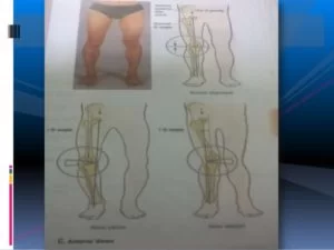
*Deformities of the knee joint:
*Genum valgum or knock knee:
~leg may be abnormally abducted.
*Genu varum or bow knee:
~leg may be abnormally adducted.
*Causes:
~Rickets
~Posture
~congenital abnormality
*Clinical features:
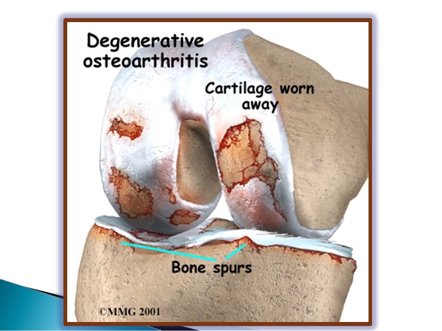
*Osteoarthritis:
~a chronic inflammatory joint disorder in which there is progressive softening & destruction of the articular cartilage, accompanied by new growth of cartilage and bone at the joint margins and capsular fibrosis.
leading to bone exposure and severe pain.
~Osteoarthritis of the knee joint is the most common joint disease.
~the knee is the most common site.
*Risk factors:
~age
~obesity
~family
*Predisposing factor:
~articular surface injury
~torn meniscus
~ligament instability
~preexiting deformity
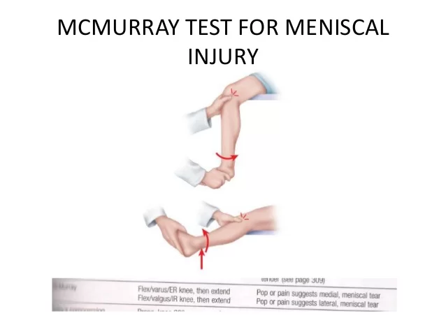
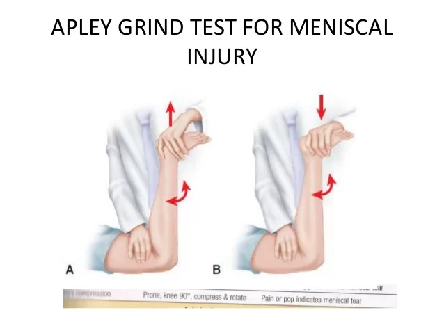
*Meniscal tear:
~ A small flap of the inner rim or the whole of the inner rim may tear.
*Causes:
~softening of menisci with advancing age.
~severe trauma.
~force-full twisting injury.
~osteoarthritis
~Diagnosis: MRI
~Treatment:-arthroscopy
*Cruciate ligament tear:
*ACL tear:
~by twisting force on a semiflexed knee.
~diagnosis:-anterior drawer test
*PCL tear:
~when an anterior aspect of the tibia is struck with the knee semiflexed so as to free the tibia backward on the femur.
~Diagnosis:-posterior drawer test
~Treatment:
~nonoperative:-robert Jones bandage
~operative: repair of ligament of reconstruction.
*collateral ligament:
~Medial collateral ligament tear:-
~by application of strong valgus force (abduction).
~detaches from its femoral attachment:-
*symptoms:
~pain and swelling on the medial side.
*Tests:
~valgus stress+30 degree knee flexion
*Lateralcollateral ligament:
~by application of strong varus force(adduction)
~detaches from its fibular attachment.
*symptoms:-pain and swelling on the lateral side.
*Tests:
varus stress +30 degree knee flexion
*Knee replacement surgery:
~in this the parts of the bone that rub together are resurfaced with metal and plastic implants.
*Component used:
~ U-shaped femoral component.
~tibial base plate
~plastic spacer/articulating surface
~patellar button.

32 Comments