
Levator Anguli Oris Muscle
Table of Contents
Levator anguli oris Muscle Anatomy
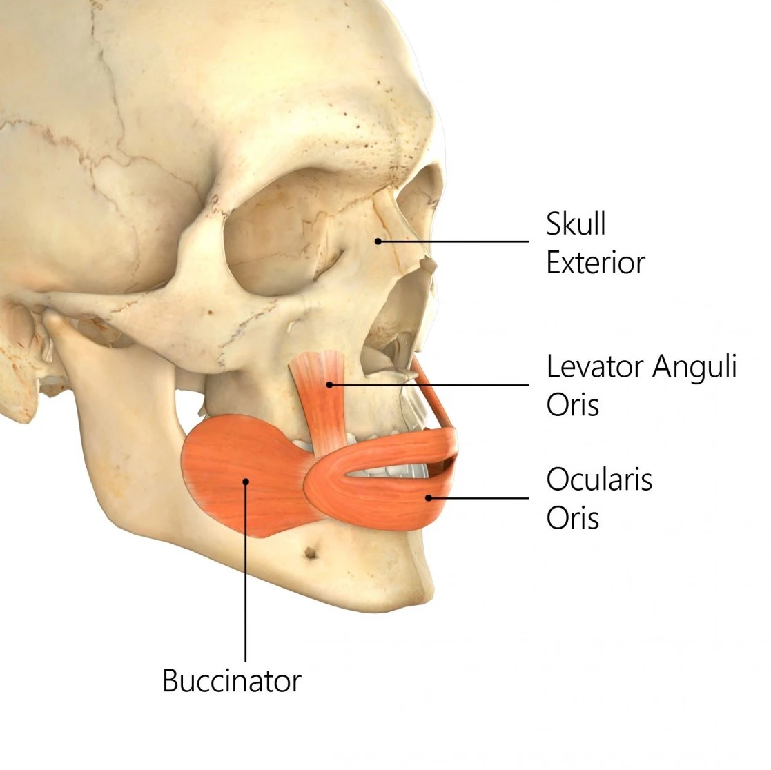
The levator anguli oris (caninus) is a facial muscle of the mouth arising from the canine fossa, immediately below the infraorbital foramen. It elevates the angle of the mouth medially. The buccal branches of the facial nerve supply the levator anguli oris.
Origin
The levator anguli oris arises from the canine fossa on the anterior surface of the maxilla right below the infraorbital foramen.
Insertion
Its fibers are inserted into the angle of the mouth, intermingling with those of the zygomaticus, triangularis, and orbicularis oris.
Nerve supply
The levator anguli oris is innervated by the buccal branches of the facial nerve.
Blood supply
Blood supply to the levator anguli oris comes from various small branches of the labial, infraorbital, and facial arteries.
Action
The levator anguli oris lifts the angle of the mouth resulting in a smile.
Contractions of this muscle produce a facial expression associated with self-confidence.
Relations
Deep and to the side of levator labii superioris is levator anguli oris. These two muscles contained the canine space, an area where odontogenic infections may spread and which is clinically relevant. On their approach to the face, the infraorbital nerve and veins travel between the levator anguli oris and levator labii superioris’ bone attachments.
Embryology
Beginning in the third or fourth week of development, the levator anguli oris muscles grow together with the other mimetic muscles of the face. The stapedial artery, which in most people later involutes, the Reichert cartilage, the stylohyoid ligament, the lesser cornu of the hyoid bone, and the styloid process of the temporal bone are all produced by the second branchial arch, which also gives rise to the muscles of facial expression.
Surgical Considerations
Even during significant facial surgery like rhytidectomy, the levator anguli oris, which is located in the medial midface, is unlikely to be iatrogenically harmed. The muscle may be in danger of damage or, at the very least, edoema and inflammation as a result of lateral rhinotomy or Weber Ferguson treatments for midfacial disease. Although uncommon, primary lesions of the levator anguli oris muscle have been observed and may be treated using an intraoral method. A 26-year-old man who presented with increasing swelling of the right cheek was found to have an intramuscular hemangioma within the levator anguli oris muscle by Koltsidopoulos and colleagues in 2013.
The levator anguli oris, which is deep to the other facial expression muscles along with the mentalis and buccinator muscles, is innervated from its deep surface. When doing procedures in the medial midface, it’s important to keep in mind how the terminal facial nerve branches relate to these muscles in order to avoid unintentionally denervating the levator anguli oris muscle. Dissecting between the levator anguli oris and the zygomaticus minor muscle is a rare surgical technique.
The levator anguli oris has been suggested as a target for midfacial rejuvenation treatments, namely plication via an intraoral incision to raise the corner of the mouth, due to its role in preserving the oral commissure’s resting posture. The treatment of flaccid facial paralysis may also benefit from this procedure. The levator anguli oris and depressor anguli oris are ideal tissue donors for lip defects, especially since sphincter tone can be maintained with the transfer of innervated muscle. When having reconstructive or lip-repositioning surgery, the levator anguli oris muscle is also a consideration.
Denewer et al. have documented utilizing the levator anguli oris muscle as a pedicle for regional flap transfer into nasal deformities in another reconstructive application. Using their method, the flap is raised by making an incision in the nasolabial fold that includes the skin, the inferior part of the levator anguli oris muscle, and, if required, intraoral mucosa from the gingivalabial vestibule. The muscle can be tunnelled into an ipsilateral nasal defect while still being linked superiorly to the maxilla.
In 1990, Takano et al. also reported the removal of the maxillary sinus’ anterior wall as part of a Caldwell-Luc technique, leaving the bone pedicled on the muscle to promote postoperative healing and showing the use of the levator anguli oris as a blood supply.
Related pathology
Facial palsy.
FAQs
The levator anguli oris is a muscle of facial expression that is responsible for elevating the angle of the mouth. When this muscle contracts, it pulls the corners of the mouth upwards, creating a smiling expression. The levator anguli oris also helps to deepen the nasolabial folds, which are the lines that run from the nose to the corners of the mouth.
The levator anguli oris, also known as the musculus levator anguli oris in Latin, is a facial muscle that is situated just above the top lip and next to the mouth’s entrance. The mouth angle is raised by the levator anguli oris.
The buccolabial muscle, or levator anguli oris, is a subdivision of the facial muscles. It is sometimes referred to as the caninus or triangularis labii superioris muscle.
References
Dao, D. D. (2023, March 17). Anatomy, head and neck: eye levator anguli oris muscle. StatPearls – NCBI Bookshelf. https://www.ncbi.nlm.nih.gov/books/NBK547688/
One Comment