
Orbicularis Oculi Muscle
Table of Contents
Orbicularis Oculi Muscle Anatomy
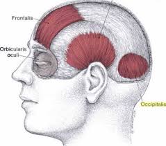
The orbicularis oculi muscle lies directly underneath the surface of the skin, around the eyes. Its function is to close the eyelid and to help in the passing and draining of tears through the punctum, canaliculi, and lacrimal sac, all parts of the tear drainage system.
Origin
The orbicularis oculi originates from the front surface of the orbital margin, which is the rim of the eye socket.
Insertion
The lateral palpebral raphe is located on the outer part of each eye socket.
Nerve supply
The upper half of the orbicularis oculi muscle receives its innervation from the temporal branch of the seventh cranial nerve (facial nerve), while the lower half receives its innervation from the zygomatic branch of the seventh cranial nerve (facial nerve).
Blood supply:
The orbicularis oculi muscle receives its blood from the branches of the facial artery and superficial temporal artery which are branches of the external carotid artery, as well as the ophthalmic artery which is a branch of the internal carotid artery.
Muscle action
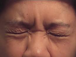
It closes lids tightly resulting in a winking, it also protects the eye from bright light. It closes lids gently resulting in blinking.
Strengthening Exercise
1. Lid Squeeze Exercise
- Sit or stand with your feet about shoulder-width apart.
- Close your eyes and place the heels of your hands over your eyes and pressed against your eyelids with your fingers resting against your forehead.
- Inhale and tightly squeeze your eyes closed as hard as you can while feeling your eyelids working to overcome the resistance of your hands.
- Hold for five seconds and relax. Repeat 10 times.
2. Surprise Exercise
- When you’re surprised, your eyelids and eyebrows raise without your knowing it.
- Sit in a comfortable chair with your feet together and your shoulders directly over your hips.
- Raise your eyebrows and eyelids as far as you can as if you were trying to touch the top of your head with your eyelids and eyebrows.
- Hold this position for five seconds and release the contraction. Repeat 10 times.
3. Look Around Exercise
- Sit or stand and look up as far as you can while keeping your head as still as possible.
- Hold this position for five seconds and then look down as far as you can without moving your head.
- Hold this position for five seconds and repeat while looking first to the left, then to the right.
- If necessary, you can repeat the entire cycle with your eyes closed as well.
4. The Partial Wink Exercise
- You can perform the partial wink exercise while standing or seated just about anywhere that you have a few minutes of free time.
- This exercise is performed by partially winking one eye at a time and holding the position for a couple of seconds.
- Avoid completely closing your eyes, since this exercise is designed to help you gain more control over your orbicularis oculi muscles.
- Try to perform two sets of 50 repetitions with each eye
Clinical Importance
The eyelids cannot close properly if the orbicularis oculi muscle is paralyzed for any reason, such as in Bell’s palsy or seventh cranial nerve paralysis. To prevent exposure to keratitis, this condition necessitates rigorous ocular lubrication with artificial tear drops and ointment. Inadequate eyelid closure can lead to severe pain, corneal ulcers, scarring, and eventually corneal perforation, which may result in the loss of the eye if left untreated.
Benign essential blepharospasm is another condition in which the orbicularis oculi muscles contract uncontrollably. Due to their inability to hold their eyes open for optimal vision, this can significantly impair their ability to do daily tasks like reading and driving. Botulinum toxin injections administered periodically to the orbicularis oculi muscle can considerably reduce symptoms.
It has an impact on the Blink Reflex, which is a component of the evaluation of the facial nerve’s functionality. Even in unconscious individuals, the facial nerve can be examined with this method.
FAQs
The muscle known as the orbicularis oculi is a sphincter that controls the eyelids. It is a wide, flat muscle that extends to three areas. A sphincter muscle contracts all the way around.
There are three parts to the orbicularis oculi: the orbital, palpebral, and deep palpebral. The muscle connects the soft tissue structures of the periorbital area to the three bones of the viscerocranium—the frontal, maxilla, and lacrimal bones.
An orbital muscle involved in facial expression is the orbicularis oculi. It is essential for shutting the eyelids and shielding the cornea from harm. Attachments: comes from the lacrimal bone, the medial palpebral ligament, and the medial orbital border.
The lacrimal bone, frontal process of the maxilla, medial palpebral ligament, and nasal section of the frontal bone are the origins of the orbicularis oculi. The superior and inferior tarsal plates, sometimes referred to as the eyelids, and the lateral palpebral raphe are where this muscle inserts.
6 Comments