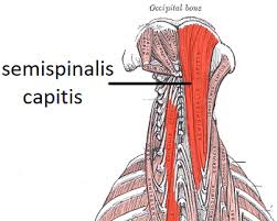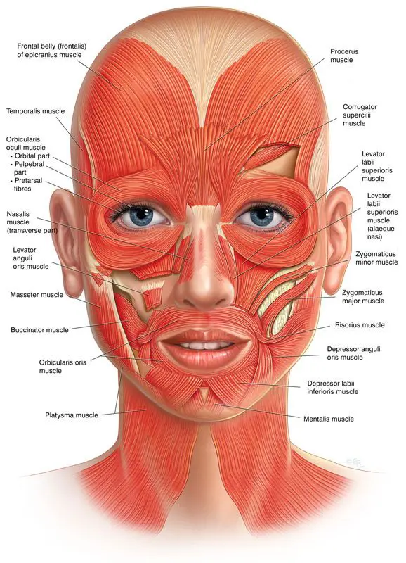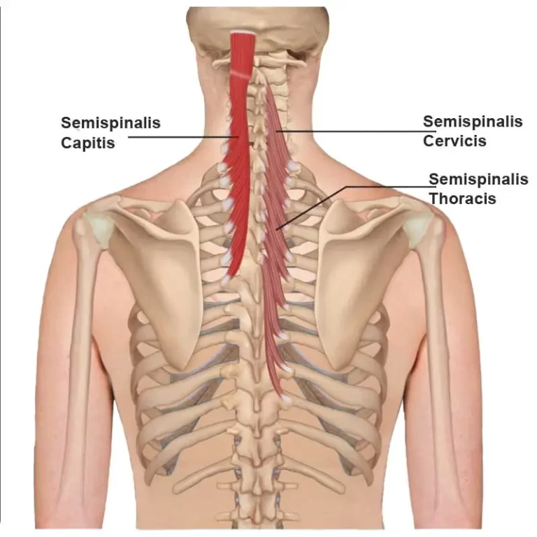
Semispinalis Thoracis
Introduction The semispinalis thoracis muscle contains thin, narrow, fleshy fasciculi, interposed between tendons of considerable length. Semispinalis thoracis muscle consists of five fascicles bridging over five to six vertebral levels between the transverse and spinous processes of certain cervical vertebrae and thoracic vertebrae. With these attachments, semispinalis thoracis muscle aids several movements of the vertebral…