Rectus capitis lateralis muscle
The Rectus Capitis Lateralis is one of the small suboccipital muscles located in the upper neck region at the back of the head. It is part of a group of four muscles known as the suboccipital muscles. These muscles are responsible for various movements and stabilizing the head and neck.
Table of Contents
Rectus capitis lateralis muscle Anatomy
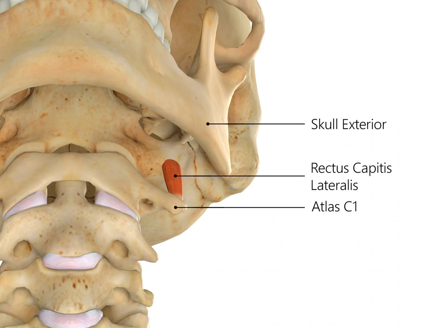
The rectus capitis lateralis (also rectus capitis lateralis muscle, lateral rectus capitis, latin: musculus rectus capitis lateralis) is a short muscle situated anterior to the vertebral column that extends between the atlas and the base of the skull and is involved in lateral flexion of the neck. Along with the rectus capitis anterior it also belongs to the anterior suboccipital muscles.
a short, flat muscle, arises from the upper surface of the transverse process of the atlas, and is inserted into the undersurface of the jugular process of the occipital bone.
Origin
Upper surface of the transverse process of the atlas.
Insertion
Under the surface of the jugular process of the occipital bone.
Nerve Supply
Anterior primary rami of the first cervical spinal nerve (C1).
Blood Supply
The muscle receives blood from the ascending cervical artery, which is a small branch of the inferior thyroid artery from the thyrocervical trunk of the subclavian artery. It also receives blood from the muscular branches of the vertebral artery. The muscle also receives small muscular branches from the occipital artery as it passes it lateral aspect.
Actions

- Lateral flexion, stabilise atlanto-occipital joint.
- Stabilizes the head
- Weakly assists with lateral flexion of the head
Clinical Importance
Neurosurgeons may use the rectus capitis lateralis muscles as a climacteric while approaching the jugular foramen. Early on in the dissection, it enables the surgeons to identify significant neurovascular structures. This becomes especially crucial if abnormal events, such tumors, have altered the native anatomy.
Along with any soft-tissue contusions, some of the most frequent injuries suffered by sportsmen are significant neck muscle strains and sprains. Strains are muscular injuries that take place whenever a muscle is overextended or stretched. Sprains occur when the ligaments or capsular structures that hold the cervical facet joints or the vertebrae together sustain damage. Because it may be difficult to distinguish between the 2 injuries, they frequently happen simultaneously. Neck stiffness, muscular spasms in the affected area, pain, or a restricted range of motion in the head or neck are all possible explanations for symptoms.
If this muscle is injured, it is treated by applying ice to the affected area, restricting movement, resting, and then receiving massage therapy. One might expect that non-steroidal anti-inflammatory medicines (NSAIDs), like ibuprofen, will treat pain or reduce inflammation. According to some reports, the rectus capitis lateralis muscles suffer an injury at a far faster rate than the other 3 pairs of muscles. The anterior muscles, or the rectus capitis lateralis, had greater accepting stiffness than the other muscle pairs.