
Semimembranosus Muscle: Origin, Insertion, Action, Exercise
Table of Contents
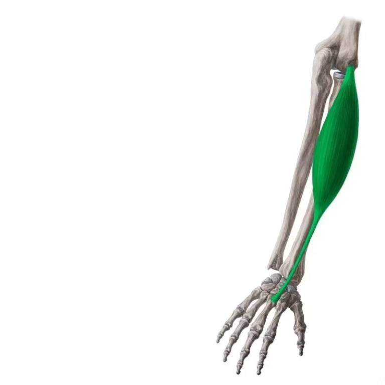
Semimembranosus Muscle Anatomy
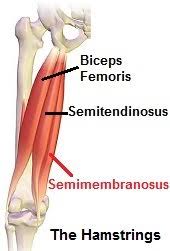
The semimembranosus muscle is one of the three muscles from the hamstring muscles in the posterior compartment of the thigh, located most medial, deep with the semitendinosus muscle in the medial side of the posterior thigh.
The other two muscles of the Hamstring group are the semitendinosus, and biceps femoris. It is a relatively long, flattened membranous muscle that spans the full length of the thigh – from the hip to the knee.
Origin:
Ischial tuberosity.
Insertion:
Medial condyle of tibia, Posterior joint capsule.
Nerve:
Tibial part of the sciatic nerve (L5, S1, and S2).
Blood Supply:
Perforating branches of femoral and popliteal arteries
Actions:
Extension of Hip and Flexion of Knee.
Function of the Semimembranosus Muscle
Because the semimembranosus muscle spans the hip and knee joints, it is accountable for a variety of joint movements. On the other hand, semimembranosus performs its role in collaboration with the other hamstring muscles.
The semimembranosus pulls the hips to extend, which straightens the upper torso when the feet are firmly placed on the ground. Internal rotation of the thigh can also be caused by semimembranosus and semitendinosus when the hip is fully stretched.
Flexion of the knee and internal rotation of the leg on the thigh result from having the legs suspended off the ground.
Standing symmetrically causes the semimembranosus and other posterior thigh muscles to become passive. But when someone leans too far forward, the semimembranosus contracts, preventing further forward motion and stabilizing the hip.
Antagonist: Quadriceps muscle.
A variant of muscles:
- Rarely absent muscle
- accessory semimembranosus muscle
Exercise of The semimembranosus muscle:
Exercise of The semimembranosus muscle is mainly Stretching exercise and strengthening exercises.
Stretching exercise:
Banded hamstring curl:
This exercise strengthens the Hamstring muscle and also the semimembranosus muscle.
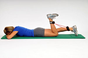
How to Perform?
- Take a prone lying position on a soft mat, with resistance around your both ankle joints.
- Your both leg should be extended behind you, toes are not tucked.
- It’s a starting position for a banded hamstring curl.
- Now breathe in and breathe out tuck in the toes of the left foot and elevated your heel towards the glutes.
- Lower down your left foot towards the mat into starting phase.
- Repeat this exercise for 10 to 15 repetitions.
- Gradually increase repetitions and sets as you improve.
Strengthening exercise:
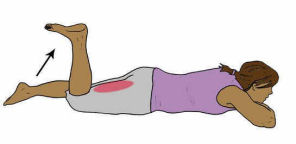
Standing Toe Touch:
How to do it?
To do this stretch you have to stand with feet and hip-width apart, then gradually bend forward from your hips and try to reach to touch your toes while keeping your knees straight. It is ok if you do not touch the ground, try to go as much as possible until you feel a slight stretch in your back thigh. when you breathe in your body takes lower down when you up to breathe out.
Structure
Interesting structural characteristics of the semimembranosus aid in the general identification of the muscle. The muscle starts off as a flat, membrane-covered structure and, halfway down the thigh, develops a fleshy belly.
Its tendon is medially connected to the fleshy component, and the fibers are directed inferomedially.
In fact, the tendon trifurcates distally to produce:
- the primary portion that attaches to the medial tibial condyle
- the popliteal fascia-fusing portion in second place
- the last portion that develops into the oblique popliteal ligament.
The fact that the semimembranosus’s lateral border forms the popliteal fossa’s superomedial wall is another significant characteristic.
Relations
Along its path, the semimembranosus muscle is surrounded by a multitude of neighboring muscular and neurovascular structures. The semimembranosus extends medially to the biceps femoris, superficially to the adductor magnus, and deep to the semitendinosus. The gluteus maximus and adductor minimus cover the muscle’s proximal region. Semimembranosus inserts on the medial tibial condyle after crossing over and becoming medially connected to the gastrocnemius medial head. The lower limb’s vessels are located in the adductor canal, which is medial to the distal part of the semimembranosus.
Around the semimembranosus tendon is a U-shaped bursa. It divides the tendon from the medial cruciate ligament, semitendinosus, medial head of the gastrocnemius, and medial tibial plateau.
Traveling lateral to semimembranosus, the sciatic nerve—the biggest nerve in the human body—ends at the apex of the popliteal fossa. At this location, the semimembranosus is partially covered and lateral to the popliteal artery and vein.
Assessment
Palpation
Although difficult to palpate, semimembranous tissue is located deep to the semitendinosus and is easier to feel when the knee is flexed.
Test of Strength
The patient lies prone with their knee extended while their strength is assessed. While the terapist applies resistance via the distal tibia and fibula in the opposite direction of flexion, the patient voluntarily flexes the knee across its range. More specifically, it can be helpful to combine the knee bend while maintaining the tibia in internal rotation for the semimembranosus.
Clinical Importance
Semimembranosus Tendinopathy
Due to its lack of knowledge, semimembranosus tendinopathy—a potential cause of chronic knee pain—often remains undetected.
This pathology may arise from age-related deterioration in elderly people or from overuse injuries sustained by marathon runners and cyclists. Patients may report mild pain in the posteromedial region of the knee that radiates either proximally to the posteromedial thigh or distally to the medial calf. Activities that involve bending of the knee exacerbate the pain.
During a clinical examination, the discomfort can be reproduced by palpating the semimembranosus tendon insertion and applying internal rotation and flexion to the knee. With rest, analgesics, and physical therapy, the majority of these patients can be treated conservatively.
Semimembranosus Muscle Pain
Pain in the semimembranosus muscle can occur due to various reasons such as overuse, strain, or injury during physical activity. This pain can range from mild discomfort to severe and can affect mobility and daily activities. Proper rest, stretching, and targeted exercises are commonly used to alleviate semimembranosus muscle pain.
Semimembranosus Muscle Injury
Damage or strain to the semimembranosus muscle, one of the three hamstring muscles at the back of the leg, is referred to as an injury. Running and jumping are two common activities that cause these injuries because they involve abrupt acceleration, deceleration, or severe muscle strain. Pain, edema, and restricted range of motion in the affected area are common symptoms.
In order to promote healing and restore function, physical therapy exercises are frequently combined with rest, ice, compression, and elevation (RICE) as part of treatment. In severe circumstances, medical treatment including medication or surgery could be necessary.
Hamstring Syndrome
Athletes with this disease frequently exhibit localized pain near the ischial tuberosity. The etiology is believed to be an ischium-related insertional tendopathy, although sciatic nerve compression may also be involved. When the hamstrings are in stress, the pain in hamstring syndrome worsens and travels down the posterior thigh or popliteal area.
This is frequently observed in hurdlers or sprinters. Upon inspection, the ischial tuberosity exhibits exquisite tenderness, and applying pressure to that area can replicate the sciatic distribution of pain. Steroid injections, anti-inflammatory medications, and rest are all part of the treatment.
Baker’s cyst
Sometimes, swollen fluid might increase the bursae that divide the muscle from the medial heads of the tibia and the gastrocnemius. This swelling is known as a “Baker’s cyst,” which was first identified as a cystic mass in children’s popliteal fossae by Morrant Baker in the 19th century.
6 Comments