Styloglossus Muscle
Table of Contents
Styloglossus Muscle Anatomy
The styloglossus muscle is one of the intrinsic muscles of the tongue, playing a role in the movement and positioning of the tongue. It is part of a group of muscles responsible for the fine motor control of the tongue’s movements.
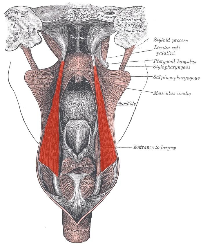
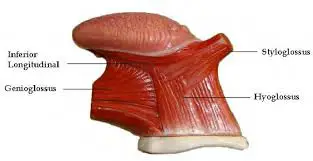
The Styloglossus muscle is the shortest and smallest of the three styloid muscles arising from the styloid process’s anterior and lateral surfaces near its apex and from the stylomandibular ligament.
The anterior and posterior bundles of the styloglossus muscle’s fibers can be separated. The bundles run bilaterally anteriorly and converge to form an arch in the midline of the tongue’s floor. The styloglossus muscle’s posterior bundles divide into around ten smaller fiber bundles that enter the tongue. These smaller bundles eventually insert into the lingual septum after passing via the genioglossus muscle and the inferior longitudinal intrinsic muscles of the tongue.
Origin:
Styloid process of the temporal bone.
Between the superior and middle pharyngeal constrictor muscles, as well as between the internal and external carotid arteries, it crosses anterioinferiorly from its origin to its insertion.
Insertion:
tip and sides of the tongue.
Nerve supply:
Hypoglossal nerve.
Blood Supply
The lingual artery’s sublingual branch supplies blood to the styloglossus muscle. The lingual artery originates at the level of the larger horn of the hyoid bone and is a direct branch of the external carotid artery. The lingual artery then passes between the deep middle pharyngeal constrictor and the superficial hyoglossus to deliver blood to the tongue. The base of the tongue is supplied by dorsal lingual arteries, whereas the body of the tongue is supplied by deep lingual arteries.
Lymphatic Drainage
The tongue is venously drained by one dorsal lingual vein and deep lingual veins. These veins empty into the sublingual vein, which drains into the internal jugular vein.
The submental and submandibular lymph nodes, which empty into the deep cervical chain of lymph nodes, receive lymphatic drainage of the front two-thirds of the tongue. The deep cervical lymph nodes get the direct drainage of the posterior portion of the tongue.
Action:
Elevates and retracts the tongue.
The tongue’s sides are drawn by the styloglossus to form an opening for swallowing. They also assist in tongue retractions since they are bilaterally active, meaning that both styloglossus muscles contract at the same time.
Assessment
The hypoglossal (XII) nerve innervates the styloglossus muscle, which is an extrinsic muscle of the tongue. Thus, by evaluating tongue motions, one may evaluate the hypoglossal nerve’s function. Patients are taught to stick out their tongues during examinations. The tongue will ipsilaterally deviate, or shift to the side of the lesion or injury when the hypoglossal nerve is damaged.
Embryology
From an embryological perspective, the development of the tongue starts in the fourth week of pregnancy, right after the pharyngeal arches appear. Every one of the initial four pharyngeal arches contributes to the act of tongue formation.
Mesodermal occipital somites give rise to myoblasts, which are the precursors of the tongue’s muscle fibers. On the other hand, ectoderm is the source of the tongue’s stromal components.
Clinical Importance
Between the middle and superior constrictor muscles of the pharynx, the styloglossus muscle forms the buccopharyngeal gap, which links the pharyngeal and submandibular areas. This clarifies the possibility of a soft tissue infection originating in the submandibular area and spreading to the pharyngeal region and adjacent tissues. Extension into the nearby retropharyngeal spaces might ensue from this transmission; further spread into the thoracic cavity could cause mediastinitis, a rare but deadly condition.
Ludwig’s angina can result in severe tongue swelling and retropulsion of the tongue base, compromising the airway. It is a potentially deadly spreading cellulitis of the submandibular, sublingual, and submental areas. A direct infection spread to the larynx, affecting tissues like the epiglottis, might cause airway compromise and edema.
FAQ
The tongue’s paired extrinsic muscles are called styloglossus muscles. It works in combination with the tongue’s other extrinsic muscles, such as the genioglossus, palatoglossus, and hyoglossus muscles, to generate the tongue’s different motions.
The lateral borders of the tongue bend upward due to the styloglossus muscle, creating an opening that allows swallowing once food particles are masticated. The tongue can also be raised and retracted in a posterior and superior manner using the paired styloglossus muscles.
Hypoglossal Nerve
The genioglossus, styloglossus, and hyoglossus are the extrinsic muscles of the tongue that are innervated by the hypoglossal nerve, along with all of the intrinsic muscles.
The styloglossus muscle runs the length of the tongue along the lateral aspect of the tongue, starting at the distal lateral aspect of the stylohyoid bone. The paired muscle joins and expands with the fibers of other tongue muscles close to the tip of the tongue. The tongue retracts when the styloglossus contracts.