
The Cerebellum: Anatomy, Function
Table of Contents
Introduction:
~The cerebellum is the largest part of the hind brain. it is situated in the posterior cranial fossa behind the pons and medulla.
~it is an infratentorial structure that coordinates voluntary movements of the body.
Relations:
~Anteriorly:-fourth ventricle,pons and medulla
~Posteroinferiorly:-Squamous occipital bone,&
~Superiorly:-tentorium cerebelli
External features:
~the cerebellum consists of two cerebellar hemisphere that is united to each other through a median vermis.
~it has two surfaces Superior and Inferior. the superior surface is convex.
~the two hemisphere are continuous with each other on this surface.
the inferior surface shows a deep median notch called the vallecula which separates the right and left hemispheres.
~the anterior aspect of the cerebellum is marked by a deep notch in which the pons and medulla are lodged.
~posteriorly, there is a narrow and deep notch in which the flax cerebelli lies.
~each hemisphere is divided into 3 lobes. the anterior lobe lies on the anterior part of the superior surface.it is separated from the middle lobe by the fissura prima.
~the middle lobe (known as the posterior lobe)is the largest of the 3 lobes.it is limited in front by the fissura prima(on the superior surface), and by the posterolateral fissure (on the inferior surface).
~the flocculonodular lobe is the smallest lobe of the cerebellum. it lies on the inferior surface, in front of the posterolateral fissure.
Subdivisions of Cerebellum:
~the cerebellum is subdivided into numerous small parts by fissures. each fissure cuts the vermis and both hemispheres. out of the numerous fissures, following the worth remembering.
1. the horizontal fissure separates the superior surface from the inferior surface.
2. the primary fissure separates the anterior lobe from the middle lobe on the superior surface of the cerebellum.
3. the osterolateral fissure separates the middle lobe from the flocculonodular lobe on the inferior surface.
The parts of the vemis are:
1.lingula
2.central lobule
3.culmen
4.declive
5.folium
6.tuber
7.pyramid
8.uvula
9.nodule
The subdivisions of the cerebellar hemisphere are:-
1.ala
2,qaudrangular lobule
3.simple lobule
4.superior semilunar lobule
5.inferior semilunar lobule
6.biventral lobule
7.tonsil
8.flocculus
Internal structure:
~outer gray matter called the cerebral cortex, extensively folded forming folia.
~inner white matter, showing distinctive tree-like pattern called arbor vitae(tree of life)
~4 pairs of nuclei white matter, the deep cerebral nuclei.
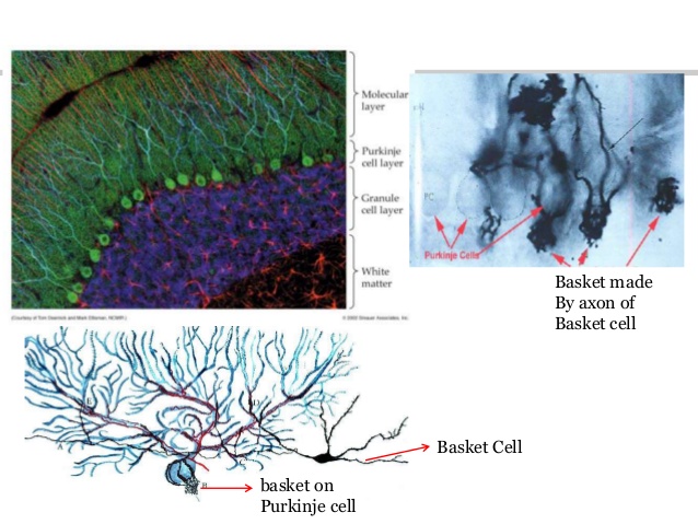
The cerebral cortex:
~microscopically composed of 3 layers,
~outer molecular
~middle,Purkinje cell layer
~inner, granule cell layer
contains 5 types of neurons:
~basket cell } molecular
~stellate cells } layer
~purkinje cell }purkinje cell layer
~granule cell }granule cell
~golgi cell }layer
~purkinje cell axons, are the only output from the cerebellar cortex, to the deep nuclei.
Input & output of the cerebral cortex:
Input:
~climbing fibers-(+,excitatory,from inferior olive ncleus)
~mossy fiber(+,excitatory,from spinal cord & brain stem)
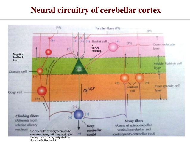
Output:
~purkinje:-(-,inhibitory)
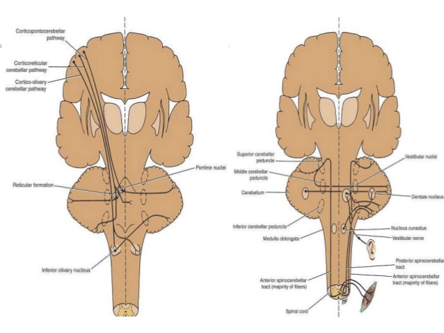
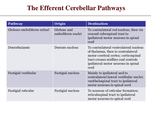
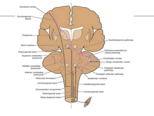
The efferent cerebellar pathways:
~the entire output of the cerebellar cortex is through the axons of the purkinje cells.
~the axons of the purkinje cells end by synapsing on the neurons of the deep cerebellar nuclei.
~the axons of the neurons from the cerebellar nuclei constitute the efferent outflow from the cerebellum.
~a few purkinje cell axons pass directly out of the cerebellum to the lateral vestibular nucleus.
~the efferent fibers from the cerebellum connect with the red nucleus,thalamus,vestibular complx,and reticular formation.
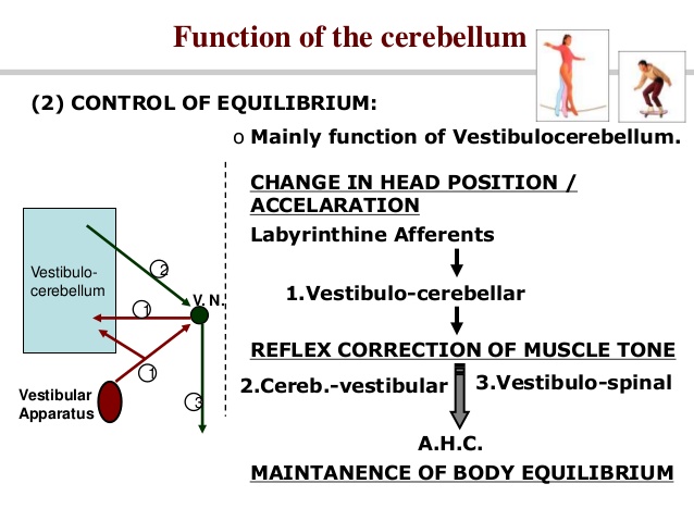
Functions of Cerebellum:
Control of tone & posture:
~mainly function of spinocerebellum:-
~change in body posture
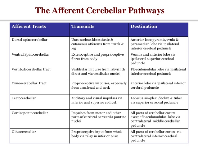
~proprioceptive from the body:-
1.spinocerebellar (dor & vent)
2.cuneo-cerebellar
3.tecto-cerebellar (visual & auditory)
~reflex correction of muscle tone:-
4.cereb-vestibular
5.cereb-reticular
6.vestibulo-spinal
7.reticulo-spinal
~A.H.C-easy maintenance of new posture.
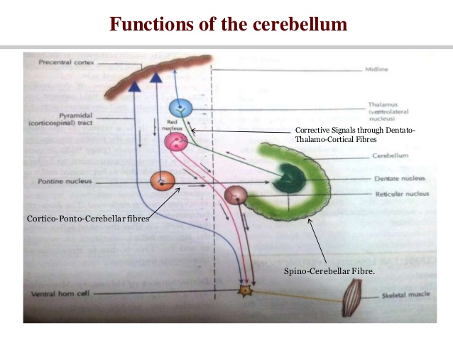
control of voluntary movement:
~act as a comparator of a servo mechanism.
~cerebellum receives two types of information.
~intended plan of movement (direct information from the motor cortex)
~what actual movements result(feedback from the periphery)
~these two are compared:-an error is calculated
~corrective output signals goes to
~motor cortex via thalamus
~brain stem nuclei and then down to the anterior horn cell through extrapyramidal tracts.
Learning new skilled voluntary movements:
~role of inferior olivery nuclei -climbing fibers
~each purkinje cell recevies input from a single climbing fibers.
~each climbing fiber cause a burst of spikes in purkinje cell (called a complex spike)when new skilled movement is done.
~complex spike causes long-term modification of the pattern of molssy fibre input to purkinjes cell.
~climbing fibre input gets increased when new skilled movement is learned.
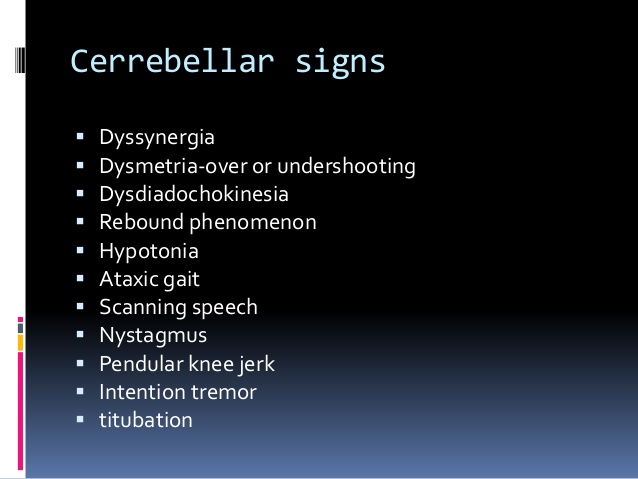
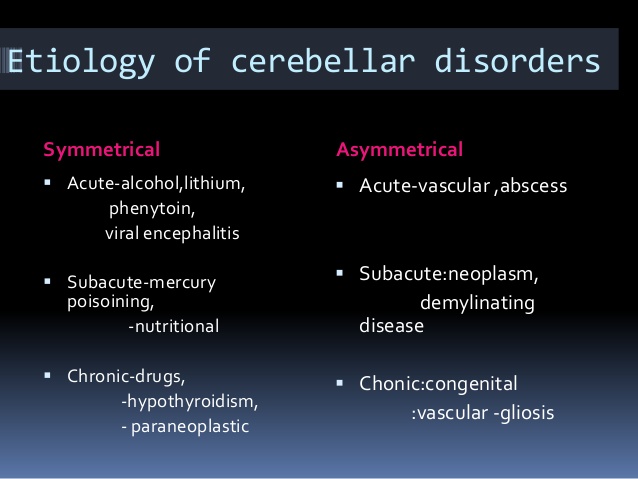
Characteristics feature:
Disturbance of posture:
~hypotonia
~face rotated towards opposite side
~body weight thrown on the healthy leg, concavity of trunk towards the affected side.
~nystagmus
~Incoordination of movement
~ decomposition of movement(movement occurring in stages)
~dysmetria:-movement is poorly carried out in direction,range,and force,past pointing(hypermetria),or falling short(hypometria)occurs.
Intention tremors:
~jerk movements O and fro on motion reaching an object.
~coarse tremor occurring at 4-6/sec.
~speech and lalling speech.
~drunken gait.
Clinical test to assess cerebellar dysfunction:
~finger nose test
~rebound phenomenon of holmes.
~a Dysdiadochokinesia-unable to carry out rapidly alternate and opposite movements.
~heel knee test
2 Comments