Scalene Anterior Muscle
Table of Contents
Anatomy of Scalene Anterior Muscle
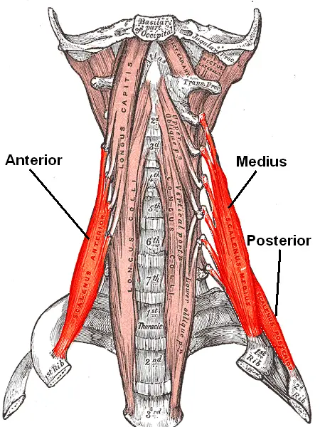
Scalene anterior Muscle is a group of three pairs of muscles in the lateral neck, namely the anterior scalene.
It is located deeply just behind the Sternocleidomastoid muscle.
The brachial plexus and subclavian artery runs between the anterior and middle scalene muscles. This provides an important anatomical location for anaesthetics to do an interscalene block.
The subclavian vein & phrenic nerve run anteriorly to the anterior scalene while the subclavian vein passes horizontally across the muscle, while the phrenic nerve runs vertically down the muscle. The subclavian artery is situated posterior to the muscle.
Origin:
Cervical vertebrae (CII-CVII).
Insertion:
First and second ribs.
Nerve supply:
Cervical nerves (C3-C8).
Blood Supply:
Ascending cervical branch of the inferior thyroid artery.
Actions:
Anterior scalene can do ipsilateral or bilaterally with the other two scalene muscles (Middle and Posterior)
Ipsilateral: flexes the neck laterally on the same side and works as a side flexor
If works Bilaterally:
If works Bilaterally contraction causes anterior flexion of the neck – Neck flexors
Ipsilateral muscle contraction creates cervical rotation to the same side – rotators
Elevate the 1st rib and that’s why it works as an accessory muscle of respiration

Elevation of first and second ribs.
Clinical Importance:
In respiratory distress, the accessory muscles of respiration come into action and the scalene muscles work as an elevation of the first two ribs to increase the lung volume. The usage of these muscles indicates difficulty in breathing.
FAQ
They function as postural muscles to keep the cervical tract in place or to actively participate in neck motions. They have the ability to tilt the neck and prevent the first rotation of the head. A neck flexion is made possible by a bilateral scalene contraction.
The anterior scalene is reached anteriorly by the subclavian vein, which crosses it horizontally, and the phrenic nerve, which descends the muscle vertically. The anterior scalene is situated anterior to the subclavian artery.
Located in the lateral region of the neck, the scalenus anterior muscle is situated behind the phrenic nerve and in front of the subclavian artery. It is associated with numerous structures of the neck; Posteriorly, it is related to the suprapleural membrane, pleura, roots of the brachial plexus, and the subclavian artery.
Grasp your SCM with your other hand’s thumb and fingers. Let go of your thumb and draw the SCM a few inches in the direction of the trapezius muscle with the other fingers.

3 Comments