Muscles Of Back Of Upper Arm
Table of Contents
What are Muscles Of Back Of Upper Arm?
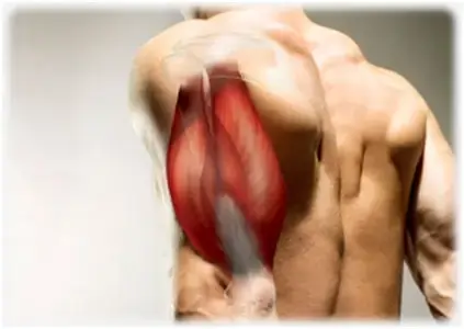
Muscles Of Back Of Upper Arm (The posterior (extensor) compartment of the upper arm) contains mainly the triceps brachii muscle. Even though the anconeus muscle is not anatomically located in the upper arm region, it is often considered to be a part of this muscle group. This is mainly because of the fact that its function is closely related to the triceps brachii muscle.
The triceps brachii muscle is the prime extensor of the forearm at the elbow joint, with help from the anconeus muscle, but is also capable of weak arm extension and adduction movements.
List of Muscles Of Back Of Upper Arm
There are two muscles which are located Back Of Upper Arm are;
- The Triceps Brachii muscles
- Anconeuus muscle
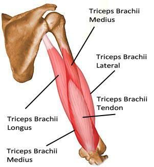
The Triceps brachii Muscle
Origin:
It arises in three heads:
The long head originates from the infraglenoid tubercle of the scapula; it is the longest of the three heads.
The lateral head originates from an oblique ridge on the upper part of the posterior surface of the humerus, corresponding to the lateral lip of the radial(spiral)groove.
The medial head originates from a large triangular area on the posterior surface of the humerus below the radial groove, as well as from the medial and lateral intermuscular septa. At the level of the radial groove of the humerus, the medial head is medial to the lateral head.
Insertion:
The long and lateral heads converge and fuse to form a superficial flattened tendon that covers the medial head and is inserted into the posterior part of the superior surface of the olecranon process. The medial head is inserted partly into the superficial tendon, and partly into the olecranon process. Although the medial head is separated from the capsule of the elbow joint by a small bursa, a few of its fibers are inserted into this part of the capsule: this prevents nipping of the capsule during the extension of the arm.
Nerve supply:
Each head of the Triceps brachii Muscle receives a separate branch from the radial nerve(C7, C8). The branches arise in the axilla and in the radial groove of the humerus.
Blood supply:
The arterial supply to the triceps brachii muscles is provided by the deep brachial artery, which is a branch of the brachial artery,
Action:
The triceps brachii muscles are a powerful extensor of the elbow. The long head of the triceps brachii muscles supports the head of the humerus in the abducted position of the arm.
Applied Anatomy:
In radial nerve injuries in the arm, the triceps usually escapes paralysis because the nerves supplying it arises in the axilla.
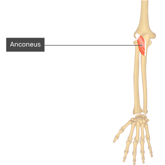
Anconeus muscle
The anconeus muscle is a small, triangular muscle on the back of the arm. The anconeus muscle is located at the posterior aspect of the elbow, extending from the distal humerus to the proximal ulna.
The anconeus muscle belongs to the superficial compartment of the extensor, along with the brachioradialis, extensor carpi radialis longus, extensor carpi radialis brevis, extensor digitorum, extensor digiti minimi, and extensor carpi ulnaris muscles.
The anconeus muscle assists in the extension of the elbow joint and provide support for both the dorsal capsule of the humeroulnar joint and the ulna itself.
Origin:
The anconeus muscle originates from a tendon on the dorsal aspect of the lateral epicondyle of the humerus bone, just proximal to the common extensor tendon. the anconeus muscles tendon lies deep in the muscle belly of the extensor carpi radialis longus and is partially attached to the dorsal capsule of the humeroulnar joint.
Insertion:
The anconeus tendon spreads out obliquely and medially into a wide muscle belly and inserts at the lateral surface of the olecranon process of the ulna and the adjoining posterior surface of the ulnar shaft. Some authors consider the anconeus muscle is a continuation of the triceps brachii muscle, due to their fibers often being partially or completely blended together.
Nerve supply:
The anconeus muscle nerve supply is the motor branch of the radial nerve, arising from root value C6-C8. The skin over the anconeus muscle is supplied by the spinal nerve T1.
Blood supply:
The Blood supply of the anconeus muscle is the recurrent interosseous branch of the posterior interosseous artery, along with contributions from the small number of musculocutaneous perforators.
Action:
Assists in the extension of the forearm at the elbow joint;
Stabilization of elbow joint
Clinical notes
It is very important to test the integrity of the radial nerve when injuries occur on/or around the arm. A proximal arm injury may completely paralyze all three heads of the triceps brachii muscle and severely limit the extension of the forearm at the elbow joint, especially against the resistance.
An injury to the midshaft of the humerus, for example, a fracture, can damage the radial nerve as it runs in the radial groove of the humerus. This injury placement may spare much of the functioning of the triceps brachii muscle and forearm extension may only be weakened, but the patient can present with a “wrist drop” due to the paralysis of muscles that extend the wrist.
Any tendon is susceptible to injury; the triceps brachii muscles tendon attachment to the olecranon process is no exception. Any activity that overuses the triceps muscle can cause the tendon to become inflamed and damaged, resulting in pain and swelling near the muscle’s attachment to the olecranon process.
Exercise of the elbow extensors muscles
Strengthening exercise of the elbow extensors muscles:
-To most efficiently use the long head of the triceps, the shoulder flexes while extending the elbow. The patient sits or stands with the elbow flexed and with a weight in the hand at the level of the shoulder. The weight is lifted straight overhead.
Caution: Good shoulder girdle muscle function is needed to effectively perform this exercise.
-Patient position sitting or standing. Begin with the long head on a stretch by positioning the humerus in Flexion with the elbow flexed, then extending the elbow against a handheld or elastic resistance.
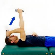
-Patient position supine with the shoulder flexed 90 degrees and the elbow flexed, with the hand either beginning by the same shoulder or the middle of the sternum(external rotation or internal rotation of the shoulder). Extend the elbow against a handheld weight or elastic resistance.
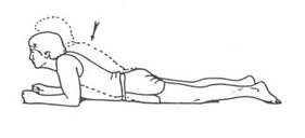
-The patient positioned is prone with the arm abducted 90 degrees and the elbow flexed over the side of the table. Extend the elbow against a handheld weight or elastic resistance.
Stretching exercise of the elbow extensor muscles:
-If the triceps is tight, use inhibition, passive stretching, and self-stretching techniques, including shoulder flexion to stretch the long head
-For self-stretching, the individual flexes the elbow and shoulder as far as possible, then pushes against the humerus with the other hand.
-To self-stretch to increase elbow flexion, the patient lies in a prone propped position with elbows flexed and forearms resting on the table. The patient lowers the chest as far as the elbow flexion allows and maintains the position as long as tolerated.