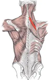
Back Of The Forearm Muscles
Table of Contents
Introduction
The muscles in the Back of the forearm muscles (posterior compartment of the forearm) are commonly known as the extensor group of muscles. The general function of these muscles is to perform extension at the wrist and fingers joints. They all are innervated by the radial nerve.
Anatomically, the muscles in the posterior compartment can be divided into two layers; the deep layer and the superficial layer. Both layers are separated by a layer of fascia.
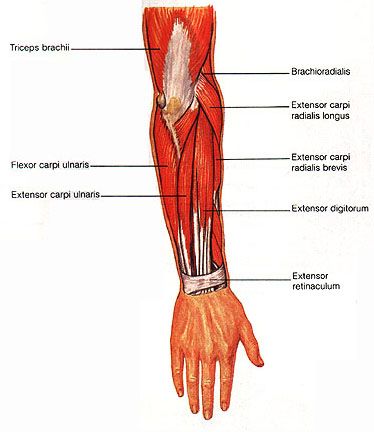
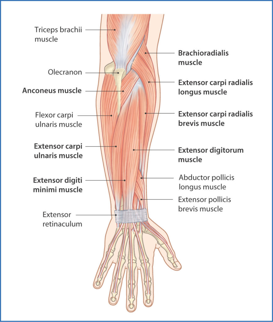
Superficial muscle of the back of the forearm
There are seven superficial muscles on the back of the forearm: They are as follows.
- Anconeus
- Brachioradialis
- Extensor carpi radialis longus
- Extensor carpi radialis brevis
- Extensor digitorum
- Extensor digiti minimi
- Extensor carpi ulnaris.
All the seven muscles cross the elbow joint. Most of them take origin (entirely or in part) from the lateral epicondyle of the humerus. This is a common extensor origin.
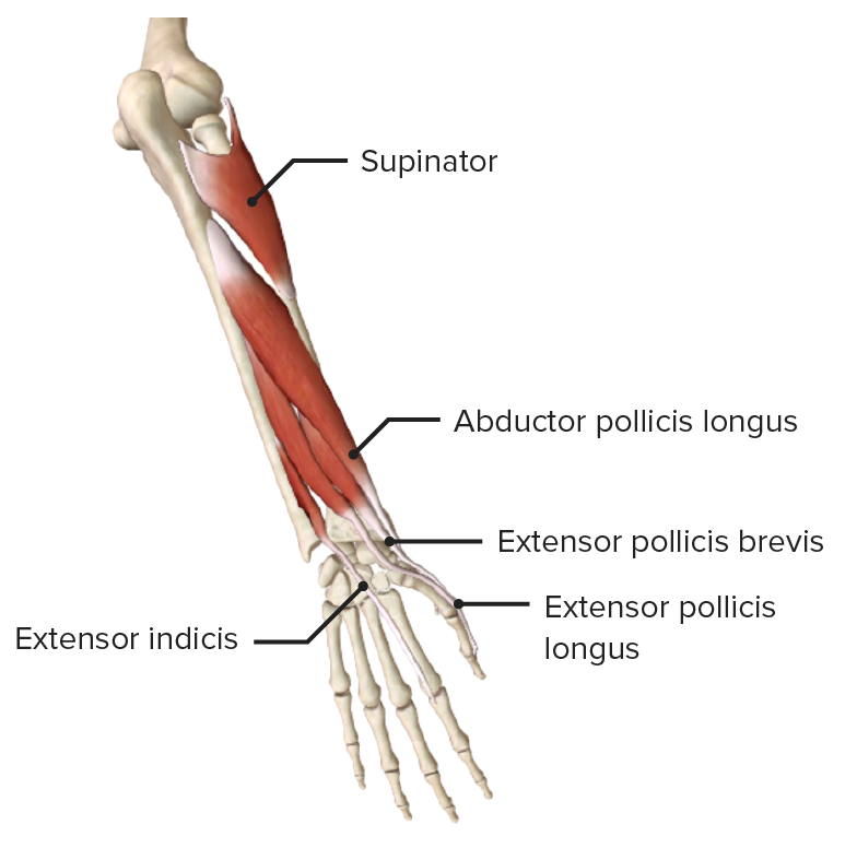
Deep muscle of the back of the forearm
These are as follows:
- Supinator
- Abductor pollicis longus
- Extensor pollicis brevis
- Extensor pollicis longus
- Extensor indicis
None of the deep muscles crosses the elbow joint. They arise from the radius, the ulna, and the interosseus membrane.
Superficial muscle of the back of the forearm
Anconeus
Origin
The Anconeus muscle originates from the posterior aspect of the Lateral epicondyle of the humerus
Insertion
The Anconeus muscle was inserted on the Lateral surface of the olecranon process of the ulna and the Upper one-fourth of the posterior surface of the ulna.
Nerve supply
The nerve supply of the Anconeus muscle is the Radial nerve(C7, C8, T1)
Blood supply
The blood supply of the Anconeus muscle is the Posterior interosseous recurrent artery
Action
Weak Extensor of the elbow joint
Brachioradialis
Origin
The Brachioradialis muscle originates from the upper two-thirds of the lateral supracondylar ridge of the humerus.
Lateral intermuscular septum.
Insertion
The Brachioradialis muscles are inserted on the lateral side of the radius just above the styloid process.
Nerve supply
The nerve supply of the Brachioradialis muscles is the Radial nerve (C5, C6, C7)
Blood supply
The blood supply of the Brachioradialis muscle is the Radial recurrent artery from the radial artery.
Action
Flexor of the forearm, especially in the mid-prone position.
Extensor carpi radialis longus
Origin
Lower one-third of the lateral supracondylar ridge of the humerus.
Some fibers arise from the common extensor origin and some fibers from the lateral intermuscular septum.
Insertion
The Extensor carpi radialis longus muscle is inserted on the Dorsum of the base of the second metacarpal bone.
Nerve supply
The nerve supply of the Extensor carpi radialis longus muscle is the Radial nerve(C6, C7)
Blood supply
The blood supply of the Extensor carpi radialis longus muscle is the Radial artery
Action
Extension of wrist
Abduction of the wrist
Assists movements of the digits by fixing the wrist joint
Extensor carpi radialis brevis
Origin
Lateral epicondyle of humerus
Radial collateral ligament of the elbow.
Insertion
The Extensor carpi radialis brevis muscle is inserted on the dorsal aspect of the bases of the second and third metacarpal bones.
Nerve supply
The nerve supply of the Extensor carpi radialis brevis muscle is the Posterior interosseous nerve (C7, C8)
Blood supply
The blood supply of the Extensor carpi radialis brevis muscles is the Radial recurrent artery, radial collateral artery, radial artery
Action
Extension of wrist
Abduction of the wrist
Assists movements of the digits by fixing the wrist joint
Extensor digitorum
Origin
The Extensor digitorum muscles originate from the Lateral epicondyle of the humerus
Insertion
The muscle ends in a tendon that splits into four parts, one for each digit other than the thumb. Over the proximal phalanx, the tendon for each digit divides into three slips one is intermediate and two are collateral. the intermediate slip is inserted on the dorsal surface of the base of the middle phalanx. the collateral slips reunite to be inserted on the dorsal aspect of the base of the distal phalanx.
Nerve supply
The nerve supply of the Extensor digitorum muscles is the Posterior interosseous nerve (C7, C8)
Blood supply
The blood supply of the Extensor digitorum muscles is the Posterior interosseous artery, radial recurrent artery, anterior interosseous artery
Action
Extension of interphalangeal, metacarpophalangeal, and wrist joints.
Extensor digiti minimi
Origin
The Extensor digiti minimi muscles originate from the Lateral epicondyle of the humerus
Insertion
The tendon joins the tendon of the extensor digitorum for the fifth digit. It is inserted through the dorsal digital expansion, into the dorsal aspect of the base of the middle phalanx, and the base of the distal phalanx.
Nerve supply
The nerve supply of the Extensor digiti minimi muscles is the Posterior interosseous nerve (C7, C8)
Blood supply
The blood supply of the Extensor digiti minimi muscles is the Radial recurrent artery from the Radial artery and the Posterior Interosseous artery from the Ulnar artery.
Action
Extention of the little finger at the interphalangeal joints and metacarpophalangeal joints.
It can help extend the wrist joint.
Extensor carpi ulnaris
Origin
Lateral epicondyle of humerus
The posterior border of the ulna
Insertion
The Extensor carpi ulnaris muscle inserted on the Medial side of the Base of the fifth metacarpal bone
Nerve supply
The nerve supply of the Extensor carpi ulnaris muscle is the Posterior interosseous nerve (C7, C8)
Blood supply
The blood supply of the extensor carpi ulnaris muscles is primarily from the ulnar artery which branches off of the brachial artery near the antecubital fossa and supplies the medial aspect of the forearm.
Action
Extension of wrist
Adduction of the hand
Fixes the wrist joint during forceful movement of the hand
Deep muscle of the back of the forearm
Supinator
Origin
The Supinator muscle originates from the Lateral epicondyle of the humerus, Radial collateral ligament of the elbow joint, annular ligament of the superior radioulnar joint, supinator crest of the ulna, and also the posterior part of the triangular area in front of it.
Insertion
The Supinator muscles are inserted on the upper one-third of the lateral surface of the radius.
Nerve supply
The nerve supply of the Supinator muscle is the Posterior interosseous nerve (C6, C7)
Blood supply
The superficial layers and deep layers of the supinator are supplied by two different sources;
The superficial layer of the supinator muscle receives blood from the radial artery, via its radial recurrent branch
The deep layer of the supinator muscle is supplied by the ulnar artery, through its posterior interosseous and posterior interosseous recurrent arteries
Action
Supination of the forearm during the elbow is in extension.
Abductor pollicis longus
Origin
The Abductor pollicis longus muscle originates from the upper part of the posterior surface of the radius and the ulna, and from the interosseus membrane.
Insertion
The tendon usually splits into two parts. One part of the split is attached to the lateral side of the base of the first metacarpal. The other part of the split is attached to the trapezium. Further fasciculi may become continuous with the opponent’s pollicis, or with the abductor pollicis Brevis muscle.
Nerve supply
The nerve supply of the Abductor pollicis longus muscle is the Posterior interosseous nerve (C7, C8)
Blood supply
The blood supply of the Abductor pollicis longus muscle is the posterior interosseous artery.
Action
Abduction and extension of the carpometacarpal joint of the thumb.
Extensor pollicis brevis
Origin
The Extensor pollicis brevis muscle is original from the Posterior surface of the radius below the origin of the abductor pollicis longus, and from the interosseus membrane.
Insertion
The Extensor pollicis brevis muscle is inserted on the dorsal surface of the base of the proximal phalanx of thumb of the thumb.
Nerve supply
The nerve supply of the Extensor pollicis brevis muscle is the Posterior interosseous nerve (C7, C8)
Blood supply
The blood supply of the Extensor pollicis brevis muscle is the posterior interosseous artery, which originates from the common interosseous branch of the ulnar artery
Action
Extension of the proximal phalanx and metacarpal joint of the thumb
Extensor pollicis longus
Origin
The Extensor pollicis longus muscle originates from the posterior surface of the shaft of the ulna and the interosseus membrane.
Insertion
The Extensor pollicis longus muscle is inserted on the Base of the distal phalanx of the thumb(dorsal aspect).
Nerve supply
The nerve supply of the Extensor pollicis longus muscle is the Posterior interosseous nerve (C7, C8).
Blood supply
The blood supply of the Extensor pollicis longus muscle is the posterior interosseous artery and perforating branches of the anterior interosseous artery
Action
Extension at all joints of the thumb.
Extensor indicis
Origin
The Extensor indicis muscle originates from the posterior surface of the ulna below the origin of the extensor pollicis longus, and from the interosseus membrane.
Insertion
The tendon joins the ulnar side of the tendon of the extensor digitorum muscle for the index finger.
Nerve supply
The nerve supply of the Extensor pollicis longus muscle is the Posterior interosseous nerve (C7, C8).
Blood supply
The blood supply of the Extensor indicis muscle is the Posterior Interosseous branch of the Ulnar artery and perforating branches of the Anterior Interosseous artery
Action
Extends the metacarpophalangeal joint of the index finger. It helps to extend the wrist joint.
Clinical significance
Intersection syndrome
Intersection syndrome is an inflammatory condition that occurs at the crossing point of the 1st dorsal compartment (Abductor Pollicis Longus muscle and Extensor Pollicis Brevis muscle ) and the 2nd dorsal compartment (Extensor Carpi Radialis Longus muscle, Extensor Carpi Radialis Brevis muscle) of the forearm.
this disease occurs due to such as rowing or weight lifting.
Intersection syndrome is best managed with rest from aggravating activities and ice or cold compresses. steroid injections may help to reduce inflammation.
Tennis elbow
Tennis elbow (or Lateral epicondylitis) is tendinopathy caused by overuse or overload of the muscles of the forearm and affects the Lateral common Extensor tendon of the elbow.
Common symptoms are pain that increases during wrist hand movements and difficulty performing day-to-day tasks.
De Quervain’s syndrome
De Quervain’s syndrome is an inflammatory condition that affects the tendon of the extensor pollicis brevis muscle and abductor pollicis longus muscles. These are the only two muscles involved in De Quervain’s syndrome.
It commonly occurs due to overuse and is referred to as one of the repetitive strain injuries. You can feel pain in the lateral side of the distal forearm, radiating to the thumb and region of the dorsal interossei.
It is usually diagnosed clinically by Finkelstein’s test, but MRI can confirm the presence of inflammation and local edema around the tendons of extensor pollicis brevis muscle and abductor pollicis longus muscle.
Exercise For Back Of The Forearm Muscles
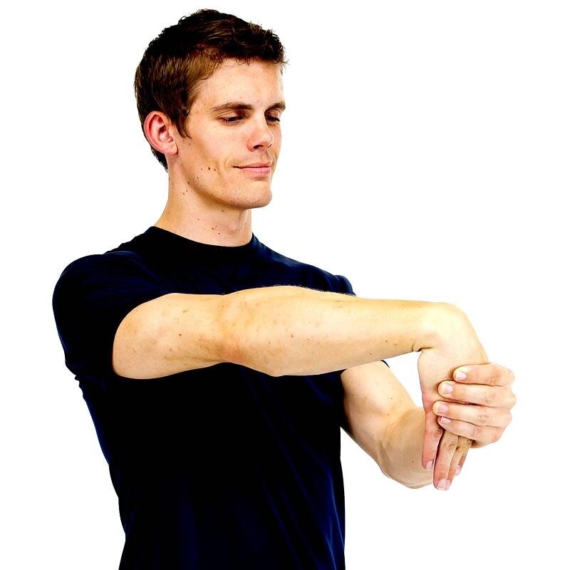
Stretching exercise
First Stretch your arm out in front of your body.
Slowly, point your fingers down until you can feel a stretch. Use the other hand to gently pull the raised hand toward your body. Hold this stretching position for 30 seconds and then relax.
Repeat this exercise three times.
Extensor stretch
Place arms at chest height with both elbows bent and the back of your hands together.
With the back of each hand touching each other, raise the wrists till you can feel a gentle stretch is felt on top of the forearm and wrists.
Hold this stretching position for 30 seconds and then relax.
Repeat this exercise three times.
Strengthening exercise
Wrist curl
Sit down comfortably with your arm resting over your knees. Hold a weight in your hand with your palms facing down and your wrist hanging over the knee.
Move your hand up as far as you can and then down as far as you can in a slow and controlled motion.
Do 10 repetitions of 3 sets.

Strengthening exercise with resistance band
Sit comfortably, resting your arm on a table with your palm facing down and your hand hanging over the edge of the table.
one end of the resistance band is put under your foot to hold it down and hold the other end in your hand. You may have to wrap it around your hand to produce some tension.
Pull up against the resistance, and extend your wrist as far as you can. Keep the motion smooth and controlled motion.
Slowly come back down to starting position.
Repeat 10 times.