Scalane Medius Muscle
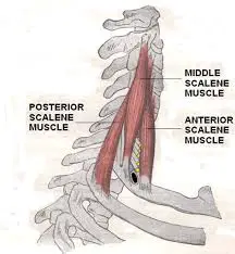
The scalenus medius muscle is one of the three scalene muscles located in the lateral neck region. It lies deeper than the anterior scalene and superficial to the posterior scalene.
Scalene medius Muscle Anatomy
The largest and longest muscle in the scalene group of lateral neck muscles is the middle scalene, also known as scalenus medius (Latin: musculus scalenus medius).
It is deeply situated, behind the sternocleidomastoid, and is frequently penetrated by the long thoracic nerve and the dorsal scapular nerve.
The posterior tubercles of the transverse processes of the lower six cervical vertebrae give rise to the middle scalene.
Origin
It originates from the posterior tubercles of C2 through C7 on the transverse process. Deviations from the origin may involve the atlas’s transverse process.
C2-C7.
Insertion
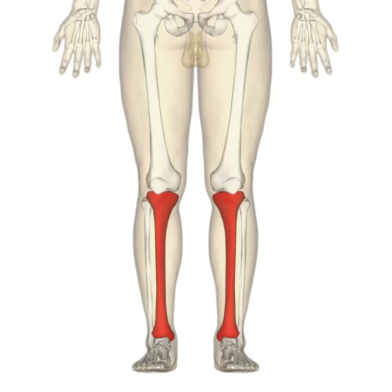
The tendon inserts to the superior border of the first rib, placing it posterior to the subclavian groove and anterior to the tubercle of the rib. One of the insertion variations is to place it onto the second rib.
Nerve supply
ventral rami of the third to eighth cervical spinal nerves.
Blood Supply
The middle scalene is supplied by the inferior thyroid artery’s ascending cervical artery branch.
Action
The middle scalenes are powerful ipsilateral flexors during unilateral contraction and collaborate with the anterior scalenes to elevate the first rib during breathing. Additionally, cervical rotation involves the middle scalene.
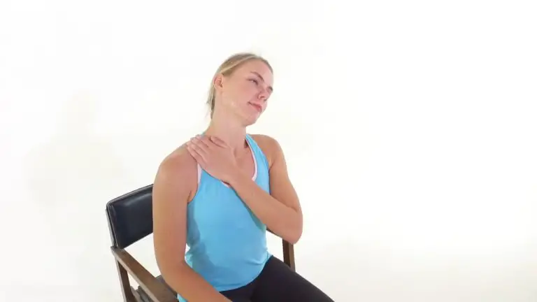
Elevate 1st rib, and rotate the neck to the opposite side.
Variations
When the middle scalene muscle inserts into the transverse processes of the atlas cervical vertebra, it may divert and end up resting on the second thoracic rib. There is a documented accessory middle scalene muscle that forms a bridge with the anterior scalene muscle in certain scientific studies. Asymptomatic pictures referring to the thoracic outlet syndrome may result from this anatomical variation.
Clinical Importance
The anatomical triangle formed by the scalenus anterior muscle (posteriorly), the scalenus medius muscle (anteriorly), and the first rib (inferiorly) are called the interscalene triangle, or scalene triangle. It is traversed by several structures, such as the brachial plexus and subclavian artery.
Thoracic outlet syndrome (TOS) can result from compression of the neurovascular structures in this area due to abnormal anatomy or injury. The ipsilateral upper limb usually has paraesthesia, pain, and weakness as a result of TOS.