Flexor Hallucis Brevis Muscle
Table of Contents
Anatomy of Flexor Hallucis Brevis Muscle
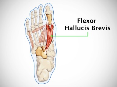
The flexor hallucis brevis muscle is a small but vital muscle located in the foot. As part of the intrinsic muscles of the foot, it plays a crucial role in maintaining proper foot function and stability during walking, running, and other weight-bearing activities.
Flexor hallucis brevis muscle is a muscle of the foot that flexes the big toe.
The plantar muscles of the foot can be categorized based on their placement into one of the four-foot muscular layers or into the lateral, medial, or central group. The flexor hallucis brevis, together with the abductor and adductor hallucis muscles, is a member of the medial compartment in the horizontal plane due to its placement. It belongs to the third layer of plantar muscles in the vertical plane, together with the flexor digiti minimi and adductor hallucis.
The tendons of the medial and lateral muscle belly that make up the flexor hallucis brevis attach to the proximal phalanx of the great toe (hallux). Two sesamoid bones form at these attachment sites and become implanted in the tendons on either side.
The great toe’s flexion at the metatarsophalangeal joint is the primary purpose of the muscles. However, the flexor hallucis brevis is also responsible for preserving the foot’s medial longitudinal arch.
Origin:
Plantar aspect of the cuneiformis, Plantar calcaneocuboid ligament, long plantar ligament.
One of the foot’s medial plantar muscles is the flexor hallucis brevis. Because the muscle originates from a bifurcate tendon, it is made up of two muscular bellies with different origins. The medial portion of the cuboid bone’s plantar surface, the lateral cuneiform bone’s neighboring surface, and the fibularis longus tendon’s groove are the sources of the lateral head. The middle band of the medial intermuscular septum and the lateral division of the tibialis posterior tendon form the origin of the medial head of the flexor hallucis brevis.
In addition, the muscle is made up of lateral and medial bellies that extend medially and anteriorly toward the great toe. Each belly’s distal tendon ends by entering the base of the hallux’s proximal phalanx on both sides. The lateral belly tendon combines with the adductor hallucis muscle during this procedure, while the medial belly tendon combines with the abductor hallucis muscle tendon.
Insertion:
- Medial Head: Medial sesamoid bone of the metatarsophalangeal joint, proximal phalanx of the great toe.
- Lateral head: Lateral sesamoid bone of the metatarsophalangeal joint, a proximal phalanx of the great toe.
Nerve supply:
the medial plantar nerve.
The medial plantar nerve (S1, S2), one of the tibial nerve’s terminal branches, innervates the flexor hallucis brevis.
Blood Supply
The flexor hallucis brevis muscle is supplied with arterial blood by the first metatarsal artery, which arises from the plantar arch’s convexity. The medial and lateral plantar arteries join to form the semicircular anastomosis known as the plantar arch. The superficial branch of the medial plantar artery, which emerges from the posterior tibial artery, also supplies blood to the flexor hallucis brevis muscle.
Actions:
flexes the proximal phalanx of the big toe.
Function of the Flexor Hallucis Brevis Muscle
The flexor hallucis brevis is primarily responsible for the great toe’s flexion at the metatarsophalangeal joint. This muscle increases the last push-off from the ground during activities like walking, running, and jumping by supporting the flexor hallucis longus during the toe-off phase of locomotion.
The flexor hallucis brevis tendons’ ability to mix with the adductor hallucis and abductor hallucis demonstrates how crucial they are for maintaining outstanding toe stability during those exercises and guaranteeing optimal force translation during the thrust phase.
By serving as a bowstring between the tarsal bones and the proximal phalanx of the hallux, the flexor hallucis brevis also contributes to the preservation of the medial longitudinal arch. The arch is raised when muscles contract, bringing the bones closer together.
Muscle Relations
Situated between the flexor digitorum brevis laterally and the abductor hallucis medially in the third layer of the medial plantar muscles of the foot, is the flexor hallucis brevis. The tendon of the flexor hallucis longus crosses between the medial and lateral muscle bellies of the flexor hallucis brevis as it runs anteromedially towards the proximal phalanx of the great toe, attaching at the base of the distal phalanx of the toe. The lateral aspect of the flexor hallucis brevis is home to the medial plantar nerve.
Within the distal tendons of the muscles, close to their attachment sites on either side of the hallux, develop two little hallux sesamoid bones. The medial and lateral tendons of the flexor hallucis brevis muscle bellies are associated with these tiny, paired, ovoid-shaped hallux sesamoid bones of the foot. They are known as the medial (tibial) and lateral (fibular) sesamoid bones of the first metatarsophalangeal joint because they are positioned on each side of the hallux. With the head of the first metatarsal articulated, the hallux sesamoid bones serve as a pivot to enhance the leverage of the flexor hallucis longus and flexor hallucis brevis.
Anatomical Variations
Origin is highly variable; fibers from the long plantar ligament or calcaneus are frequently received. attachment to the cuboid bone that is not always acceptable. Slip to the second toe’s initial phalanx.
Clinical Importance
The most common symptoms of flexor hallucis brevis dysfunction are toe malformations, pain and trouble during locomotion, and pain in the ball of the foot when extending the big toe. Sesamoiditis or FHB muscle damage could be the cause of this.
Walking or running on uneven terrain, wearing high heels, or wearing shoes that are too small can aggravate the muscle in the second toe.
Assessment
Palpation:
The FHB muscle is deep in the foot, making it almost tough to palpate. (The foot muscles’ third layer out of four layers).
Strength of muscles:
MMT grading can be used to manually measure FHB strength. The patient should begin the test with their foot hanging over the table in a supine or long sitting position. To provide stability, place your hand slightly below the ankle and instruct the patient to bend their big toe while you use your other hand’s fingers to oppose the movement.
Stretching:
Put your big toe as far into hyperextension as you can, hold it there, and then release the muscle.
Strengthening
For the purpose of strengthening the big toe and the other four toe flexors, pulling a towel is always a good idea. However, it is important to ensure that the patient is engaging the FHB muscle, which is responsible for bending the great toe at the MP joint. Using a resistance band or placing a weighted object on the towel, you can advance the activity.
FAQs
Numerous activities, such as walking, running, or even just standing on uneven or rough surfaces, can cause injuries to the flexor hallucis brevis. Additionally, injuries can result from poorly fitted shoes, especially those that are too tiny. Long-term use of high-heeled shoes can cause injuries to this muscle in women.
One of the foot’s short muscles is the flexor hallucis brevis (FHB). It splits into two halves up front, which are placed into the medial and lateral bases of the great toe’s first phalanx.
To strengthen the flexor hallucis brevis, focus on exercises like toe curls, toe scrunches, and towel pickups using your toes. These help target and strengthen the muscles in your foot.
Flexion of the proximal phalanx at the great toe’s metatarsophalangeal joint is achieved by the flexor hallucis brevis muscle. It can be examined by holding the great toe’s interphalangeal joint extended and stretching the proximal phalanx at the metatarsophalangeal joint against resistance.
The big toe, or first metatarsophalangeal joint, is flexed by the flexor hallucis brevis. It aids in keeping the medial longitudinal arch in place. It provides more push-off during the gait’s toe-off phase.

