Striated Muscle Tissue
Table of Contents
What is Striated Muscle Tissue?
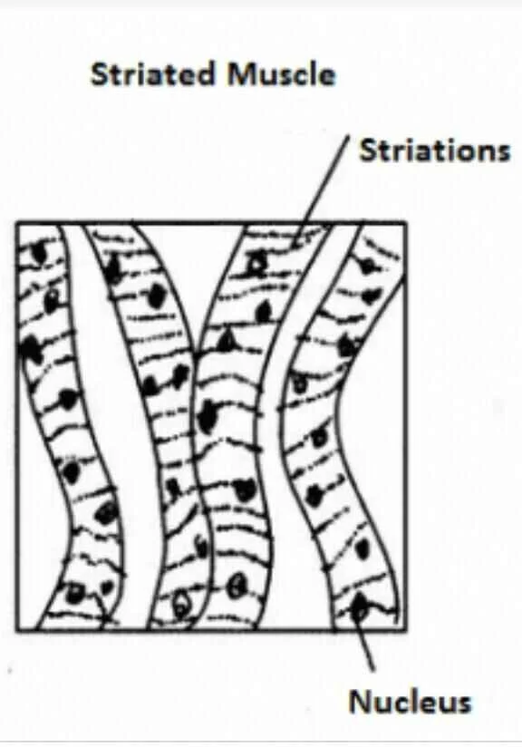
Sarcomeres, which are recurring functional units seen in muscle tissue, are characteristics of striated muscle tissue. The striated appearance seen in microscopic photographs of this tissue is caused by the presence of sarcomeres, which appear as a series of bands seen along the muscular fibers.
Two varieties of striated muscle exist:
- Cardiac muscle (heart muscle)
- Skeletal muscle (muscle attached to the skeleton)
Structure
T-tubules, which are found in striated muscle tissue, allow the sarcoplasmic reticulum to release calcium ions.
Skeletal muscle
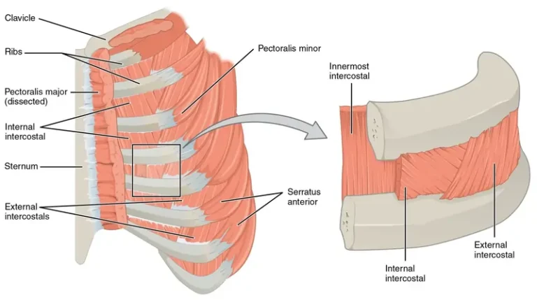
Blood vessels, nerve fibers, connective tissue, and skeletal muscle fibers are all components of skeletal muscle. The epimysium that surrounds skeletal muscle allows the muscle to maintain its structural integrity even during contractions.
The muscle fibers, covered with endomysium and collagen, are arranged into bundles by the perimysium. Sarcolemma, sarcoplasm, and sarcoplasmic reticulum are found in every muscle fiber. Sarcomeres are the functional units of muscular fibers.
Actin and myosin myofilaments are repeated as sarcomeres to form myofibrils, which are found in every muscle cell. Each muscle cell, which is arranged at regular intervals underneath the sarcolemma, has many nuclei.
Skeletal muscles can be categorized as either fast-oxidative (Type II) or slow-oxidative (Type I) based on their contractile and metabolic characteristics.
Cardiac muscle
In the heart, cardiac muscle is located between the epicardium and the endocardium. Typically, cardiac muscle cells have a single nucleus that is situated in the center. Both mitochondria and myoglobin are abundant in them.
The cells of cardiac muscle are unicellular, in contrast to skeletal muscle. Gap junctions and desmosomes are found in the intercalated discs that link these cells to one another.
Striated versus smooth muscle
Since smooth muscle tissue lacks sarcomeres, it is not striated like skeletal and cardiac muscle tissue is. Smooth muscle is found in hollow structures like the walls of blood arteries and intestines, whereas skeletal muscles are connected to some part of the skeleton.
Smooth muscular fibers are spindle-like with tapered ends, whereas striated muscle fibers are cylindrical with blunt ends. Compared to smooth muscle, striated muscle tissue has more mitochondria. Skeletal muscle cells have several nuclei, whereas cardiac and smooth muscle cells only have one.
Function
Striated muscular tissue’s primary purpose is to contract and generate force. The heart’s contractions will circulate blood throughout the body. The contractions of skeletal muscle allow for breathing, movement, and preservation of posture.
The heart’s pacemaker cells’ myogenic response causes contractions in the cardiac muscle. The autonomic nervous system sends messages to these cells, which causes the heart rate to change. Cells used in pacemakers are autorhythmic.
The heart rate is determined by the predetermined intervals at which they depolarize to threshold and fire action potentials. The pacemaker cells depolarize other cardiac muscle fibers such that they contract simultaneously because of the gap junctions.
Skeletal muscle fibers depolarize in response to signals from motor neurons, which causes the sarcoplasmic reticulum to release calcium ions. Actin and myosin filament mobility is accelerated by calcium.
The muscle contracts as a result of the sarcomere shortening. The mysia attaches to the periosteum which protects the bone in the skeletal muscles that are attached to tendons that pull on bones. Before the bone moves, the mysia, tendon, and periosteum will all be affected by the muscle’s contraction. Additionally, the mysia may attach to fascia or an aponeurosis.
Damage Repair
After an injury, adult humans are unable to repair cardiac muscle tissue, which can cause scarring and ultimately heart failure.
Small quantities of heart regeneration can be completed by mammals while they are developing. Other animals can repair heart muscle tissue for the duration of their lives.
Because satellite cells remain quiescent in all healthy skeletal muscle tissue, skeletal muscle can regenerate significantly more quickly than cardiac muscle.
Some of these processes include the inflammatory response, the maturation and remodeling of freshly generated myofibrils, and the activation, differentiation, and fusion of satellite cells. Damaged muscle fibers necrotize first in this process, which sets off the inflammatory reaction. The cell debris is phagocytosed by macrophages.
Eventually, they will release anti-inflammatory cytokines, causing the inflammation to stop. Additionally, these macrophages can promote satellite cell differentiation and proliferation. To proliferate, the satellite cells reenter the cell cycle. After that, they exit the cell cycle to either develop into myoblasts or self-renew.
Dysfunctions
Skeletal muscle
- Sarcopenia
- Polymyositis
- Dermatomyositis
- Inclusion body myositis
Cardiac muscle
- Coronary artery disease
- Arrhythmia
- Cardiomyopathy
FAQ
Since they exhibit alternating bands of light or dark, or striations, when examined under a microscope, these muscles are sometimes called striped. The cells are organized into bundles and have a long, cylindrical form. They have several nuclei and are multinucleated.
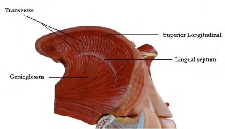
Skeletal or striped muscles are other names for striated muscles. Our throat, tongue, hands, feet, and abdomen all contain them. They are composed of cylindrical, long, and thin muscular fibers that are packed together. The sarcoplasm of these multinucleate fibers contains myofibril molecules.
Striated muscles provide the force for all other voluntary motions of the body in addition to locomotion. Unstriated or involuntary muscles are another name for smooth muscles. The long fusiform or spindle-shaped cells or fibers that make up smooth muscles are arranged in bundles or sheets.
Skeletal muscle and cardiac muscle are the two tissue types that make up striated musculature. The majority of muscles that are linked to bones are composed of skeletal muscle. The term “skeletal” was born. On the other hand, the muscle that lines the walls of the heart is called cardiac muscle.
muscle with striae. noun. : muscular tissue that is primarily controlled by voluntary action; compared to smooth muscle, voluntary muscle; composed of long, thin cells with several nuclei and alternating light and dark stripes; typically links to and moves the vertebrate skeleton.
The human body has both smooth and striated muscle; however, striated muscle is related to the skeleton, whilst smooth muscle is located in the viscera or internal organs. Smooth muscle is made up of linked cells that form layers, whereas striated muscle is made up of muscular fibers that are constructed of thick and thin filaments.
Because of their very elongated structure, these cells—which may reach enormous lengths of 2-3 cm and diameters of 100 μm in an adult human—are sometimes referred to as muscle fibers. Every one of them is a syncytium, with several nuclei sharing a single cytoplasm.
There are three distinct types of muscle tissue: skeletal, smooth, and cardiac. The heart’s walls include cardiac muscle cells, which are striated or striped and controlled involuntarily.
Because it is primarily linked to the skin and bones, the striated muscle, sometimes referred to as the skeletal muscle, is what allows the body and limbs to move.
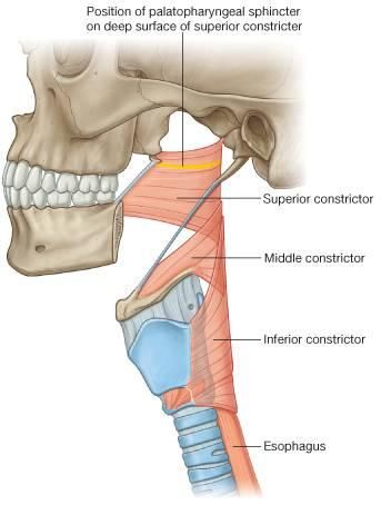
face, neck, limbs, etc. contain striated muscles. Additionally, the diaphragm, tongue, throat, and upper portion of the esophagus contain them.
The long, unbranched cells comprising striated skeletal muscle are grouped parallel in sheets and tendons. Their diameter can vary from 10 to 100 µm, and their length can be several to thirty centimeters. Their many nuclei are situated just beneath the plasma membrane.
The muscle cells in the skeleton are long, cylindrical, and striped. They have several nuclei, which is what it means to be multi-nucleated. This is a result of their formation by the merging of embryonic myoblasts. Every nucleus controls the surrounding sarcoplasm’s metabolic needs.
The heart’s walls include cardiac muscle cells, which are striated or striped and controlled involuntarily. Smooth muscle fibers are spindle-shaped, and found in the walls of all hollow visceral organs (such as the liver, pancreas, and intestines), except the heart. They are also controlled involuntarily.
The heart’s thick central layer is made up of cardiac muscle, often known as the myocardium. Together with skeletal and smooth muscle, it is one of the three different kinds of muscle in the body. An inner endocardium and a thin outer layer known as the epicardium (also known as the visceral pericardium) encircle the myocardium.