Spina bifida
Table of Contents
What is spina bifida?
- The spina bifida mean “split spine” in Latin words. Spina bifida is a birth defect in which an area of the spinal column does not form properly, leaving a section of the spinal nerves and spinal cord exposed through an opening in the back region of a newborn baby. It is a type of neural tube defect(NTD), in which the neural tube is the structure in a developing embryo that eventually becomes the baby’s spinal cord, brain, and the tissues that enclose them.
- Generally, the neural tube forms the starting phase of early pregnancy and closes by the 28th day after conception. In newborn babies with spina bifida, a portion of the neural tube does not develop or close properly, which causes problems in the spinal cord, nerves, and the bones of the spine.
- Spina bifida might cause intellectual and physical disabilities that can be varied from mild to severe, depending on the type of defect, location, size, and complications. When required, early treatment for spina bifida involves surgery, but such treatment can not always completely resolve the issue.
- Spina bifida is the most general central nervous system birth defect and occurs in 1 per 2,000 live births in the United States. Approximately 1,500 babies are born with spina bifida in the U.S. every year.
What are the types of spina bifida?
The below given three most common types of spina bifida are:
Spina bifida occulta
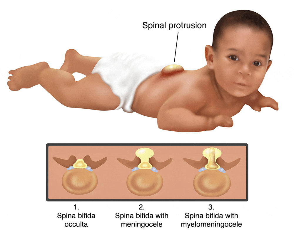
- It is also known as “hidden” spina bifida. It is the mildest and most common type of this condition. Spina bifida occulta results in a small gap or separation in one or more of the bones of the spine means vertebrae but no sac or opening on the back. In this type of spina bifida, there is no damage to the nerves or the spinal cord. Many patients who have spina bifida occulta do not even know this unless the condition is diagnosed during an imaging test done for any other reasons. Many times, spina bifida occulta is not identified until adulthood or late childhood. The nerves and the spinal cord generally are normal. It does not cause any disabilities and may not be noticed until later in life.
- It generally only includes a minimal portion of the spine, in which it generally occurs without symptoms and does not need treatment. In a few patients, the skin overlying the bony defect will show precise changes, such as a dimple, a tuft of hair, or red or purple coloring. It has been estimated that approximately 10% to 20% of the U.S. people have spina bifida occulta and most of them do not even know they have this type of condition.
- In rare cases, spina bifida occulta will cause problems when a baby develops into adolescence. By this time in the life of the child, the spinal cord has become rapidly to the backbone. When the growth spurt of adolescence begins, the nerves of the spinal cord become stretched. The result can be difficulties such as numbness and weakness in the legs, bladder infections, and incontinence means a lack of bowel and bladder control. The higher the spinal cord is stretched, the worse the symptoms of this type of spina bifida become. Surgery to relieve these symptoms by reducing the tension on the spinal cord is simple and frequently successful.
Myelomeningocele
- It is also known as open spina bifida, myelomeningocele is the most severe and dangerous type occurring nearly once for every 1,000 live births. The spinal canal is open along a few vertebrae in the middle or lower back. For babies born with a myelomeningocele, the spinal cord does not form in the proper manner and a portion or part of the undeveloped cord protrudes through the back. A sac contains cerebrospinal fluid (CSF) and blood vessels surrounding the protruding cord, which is generally not covered by the skin so that the tissues and nerves are exposed.
- Between 70% and 90% of newborns with myelomeningocele also have hydrocephalus because a defect at the base of the skull means Chiari malformation. Hydrocephalus is an excess buildup of spinal fluid on the brain that will cause seizures, brain damage, or blindness if it is not treated. To avoid this condition, plastic shunts must be surgically inserted beneath the skin to drain extra fluid into the abdominal cavity.
- Infants born with myelomeningocele frequently have weakness or paralysis below the level of the spinal lesion. This affects the lower limbs along with problems with bowel and bladder function. In extreme or serious cases, the upper extremities and trunk are involved.
- Patients with myelomeningocele have physical disabilities that can range from moderate to severe. These disabilities may include:
- Incontinence
- Difficulty going to the bathroom or toilet
- Inability to move or feel their legs or feet
Meningocele
- This rare type of spina bifida is characterized by the meninges which is the membrane surrounding the spinal cord, protruding through the opening causing a sac or lump on the back. It is generally little or no nerve damage and the spinal cord is not in the fluid sac. The baby has no neurological issues. Babies with meningocele may have a few minor disabilities with functioning, including those affecting the bowels and bladder.
- In this least general type of spina bifida, More severe than spina bifida occulta, meningocele can never be repaired by surgery with no or little nerve damage resulting.
What are the symptoms of spina bifida?
Spina bifida occulta symptoms
Generally, there are not any symptoms or signs as the spinal nerves are not involved. However, A doctor or health care provider can sometimes see signs on the newborn baby’s skin above the spinal issues, including a small dimple, a tuft of hair, or a birthmark. In some cases, these skin marks can be signs of an underlying spinal cord problem that can be identified with spinal ultrasound or an MRI in a newborn baby.
Symptoms of spina bifida occulta include:
- A gap in between vertebrae
- No fluid-filled sack outside the body
- No visible opening outside
- Small dimple or birthmark on the back
- Small cluster or group of hair on the back
- An area of extra fat on the back
- A patient may not even know they have this type of spina bifida.
Meningocele symptoms
Symptoms of meningocele type of spina bifida include:
- A small opening in the back
- Sack that can see at birth
- Normal development of the spinal cord
- Membranes pushing out through the opening in the vertebrae into the sack
- Membranes can be surgically removed in cases of meningocele.
- This type may cause problems with bowel and bladder function.
Myelomeningocele symptoms
In this severe type of spina bifida:
The spinal canal stays open along a few vertebrae in the middle or lowers back
Both the spinal nerves or cord and the membranes protrude at birth, forming a sac
nerves and tissues generally are exposed, though few times skin covers the sac
- The myelomeningocele lesion can occur at any level on the spine and is developing, but most are seen in the lumbosacral region. Depending on the lesion’s level or location, myelomeningocele may cause:
Bladder problems
Many patients with spina bifida have problems passing and storing pee. This is caused by the nerves that control the bladder not forming correctly. It can develop issues such as:
- Urinary tract infections (UTIs)
- Urinary incontinence
- Hydronephrosis – where one or both kidneys become swollen and stretched because of a build-up of pee inside the kidneys
- Kidney stones
- Kidney scarring
- The kidneys and bladder will need to be monitored regularly because of the higher risk of infection. Ultrasound scans may be required, along with that tests to measure the pressure and bladder volume inside it.
Bowel problems
The nerves that run through the spinal cord also control the sphincter muscles that keep poo in the bowel and the bowel.
Many patients with spina bifida have no or limited control over their sphincter muscles and have bowel incontinence.
Bowel incontinence frequently leads to periods of constipation followed by soiling or episodes of diarrhea.
Movement problems
Loss of sensation and weakness below the level of lesion
The brain controls all the muscles in the body with the nerves that pass through the spinal cord. Any damage or injury to the nerves can cause problems controlling the muscles of the body.
Most patients with spina bifida have some degree of paralysis or weakness in their lower limbs. They may need to use crutches or ankle supports to help them move around. If patients have severe paralysis, they may need a wheelchair.
Paralysis can also cause other associated issues. For example, as the muscles in the legs are not being used properly, they can become very weak.
The muscles support the bones, as a result, muscle weakness can also affect bone development. This can cause deformed or dislocated joints, misshapen bones, bone fractures, and abnormal curvature of the spine known as scoliosis.
Orthopedic malformation conditions such as club feet or problems of the hips or knees
Sexual dysfunction
Cognitive impairments
Skin problems
The reduced sensation can make it hard to tell when the skin on the legs is damaged, for instance, if the skin gets burnt on a radiator and a patient with spina bifida injures their legs without realizing it, the skin could become infected or an ulcer could happen. It is essential to check the skin regularly for signs and symptoms of injury.
latex allergy, patients with spina bifida can produce an allergy to latex; symptoms can range from a mild allergic reaction including, skin rashes and watery eyes, to a severe allergic reaction, known as anaphylactic shock, which requires an immediate injection or medication of adrenalin.
Spina bifida can lead to a variety of emotional and social challenges and lifelong quality of life problems.
In some cases, the brain develops an Arnold-Chiari II malformation, in which the hind-brain descends or herniates into the upper portion of the spinal canal in the neck. This herniation of the hind-brain blocks or reduces the circulation of cerebrospinal fluid, which causes hydrocephalus means accumulation of fluid in the brain, which can injure the developing brain. Ventricular shunting, which means the placement of a thin tube into the ventricles of the brain is used to drain fluid and relieve hydrocephalus.
Hydrocephalus
Many patients with spina bifida and hydrocephalus will have normal intelligence, however, few will have learning difficulties, such as:
- It can cause additional symptoms immediately after birth, such as seizures, irritability, drowsiness, poor feeding, and being sick.
- A short attention span
- Difficulty reading
- Difficulty solving problems
- Difficulty understanding some spoken language, especially fast conversations between a group of people
- Difficulty making detailed plans or organizing activities
- They may also have problems with physical and visual coordination, for example, tasks such as fastening buttons or tying shoelaces.
In some patients, the lower parts of the brain go downwards towards the spinal cord, which is known as type 2 Arnold-Chiari malformation and is linked to hydrocephalus.
Spina Bifida in Adults
Adults who have spina bifida face various issues than do children, including:
- The normal aging process includes loss of flexibility and muscle strength, low physical stamina, and a reduction in sensory
- abilities tend to decline quicker or more severely for adults with spina bifida.
- Spinal cord tethering, where the spinal cord becomes attached to surrounding tissue that causes rapidly progressing scoliosis, symptoms of skin sores, loss of sensation, pain particularly in the lower extremities or testicles, and urinary tract leakage or infections.
- Changes in bowel patterns including abdominal pain or constipation.
- Orthopedic problems such as the early onset of arthritis, osteoporosis, and progressive back pain.
- Loss of skin sensation along with poor circulation, bruising, inability to sweat, and slow wound healing.
- Latex allergy
- High blood pressure
- Central and obstructive sleep apnea can lead to long-term damage to the heart.
- Adults with this condition tend to have high rates of obesity.
- Females with spina bifida are able to get pregnant, but their condition can make pregnancy more complex.
What causes the spina bifida?
No one knows exactly what causes spina bifida. It is thought by some scientists and researchers to result from a combination of nutritional, genetic, and environmental risk factors, such as folate (vitamin B-9) deficiency in the mother’s body and a family history of neural tube defects.
But doctors and health care professionals do know that the condition is more common among Hispanic and white babies and in girls. Along with that, women who are obese or who have diabetes that is not managed well may be more likely to have a child with spina bifida.
What are the risk factors for Spina bifida?
Spina bifida is mostly seen among Hispanics and white people, and females are affected more frequently than males. However, researchers and doctors do not know for sure why spina bifida occurs, but they have identified some risk factors:
Folate deficiency
Folate, which is the natural form of vitamin B-9, is essential to the development and growth of a healthy baby. The synthetic form, found in fortified foods and supplements, is known as folic acid. A folate deficiency raises the risk of spina bifida and other neural tube defects issue.
Family history of neural tube defects
Couples who have had a child with a neural tube defect have some amount of higher chance of having a second baby with the same problem. That risk raises if two previous children have been affected by the spina bifida. Moreover, females who were born with a neural tube defect have a higher chance of giving birth to a child with this condition than someone who does not have a neural tube defect. However, most children with spina bifida are born to parents with no known family history of the condition.
Diabetes
Females with diabetes who do not have well-controlled blood sugar have a greater risk of having a baby with spina bifida.
Some medications
For instance, anti-seizure medications, like as valproic acid seem to cause neural tube defects when taken during pregnancy by the mother. This might happen because they interfere with the body’s ability to use folic acid and folate.
Increased body temperature
Some information suggests that higher body temperature means hyperthermia in the early weeks of pregnancy may raise the risk of spina bifida. Increases in core body temperature, because of fever or use of a hot tub or sauna, have been associated with some amount of increased risk of spina bifida.
Obesity
Pre-pregnancy obesity is connected with a raised risk of neural tube birth defects, including spina bifida.
If someone has known risk factors for spina bifida, talk with their doctor to determine if they need a prescription dose or a larger dose of folic acid, even before pregnancy starts.
What are the complications of spina bifida?
Spina bifida may cause minor physical difficulties or minimal symptoms. However, severe spina bifida can develop more crucial physical disabilities. Severity is affected by:
- The location and size of the neural tube defect
- Skin covers the affected area or not?
- Which number of spinal nerves pop out of the affected area of the spinal cord?
This list of possible complications may seem in higher number, but not all patients with spina bifida get all of the below-given complications. Many of given these complications can be managed.
Paralysis
Patients with paralysis caused by spina bifida will need life-long assistance from leg braces, wheelchairs, or crutches to help them move around or walk. Paralysis may be complete or partial. Complete paralysis is the total loss of function and feeling in the affected area, while partial paralysis means a partial loss of function and feeling, such as the inability to move a leg.
Walking and mobility problems
Below the area of the spina bifida defect, the nerves that control the leg muscles do not work correctly, which can cause muscle weakness in the legs and a few times paralysis. Whether a child can walk generally depends on the location of the defect, its size, and the care received after and before birth.
Orthopedic complications
- Kyphosis and scoliosis are conditions identified by abnormal curves in the spine. An increased forward rounding of the back is hunchback or kyphosis and scoliosis is an abnormal sideways curve. It is mostly seen in older women and is frequently related to osteoporosis.
- Clubfoot is another mostly seen orthopedic complication, and it is a condition in which the foot is twisted out of its normal position.
- Abnormal growth
- Dislocation of the hip
- Bone and joint deformities
- Muscle contractures
- Orthopedic issues associated with spina bifida may be caused by spinal cord tethering, unstable muscles around joints, paralysis, and reduced feeling in the feet and legs. The types of issues depend on the location of the defect.
Bowel and bladder problems
Bladder and bowel problems are mostly seen in patients with severe forms of spina bifida because the nerves that control these functions generally do not work correctly and have been damaged when patients have myelomeningocele. This is due to the nerves that supply the bladder and bowel coming from the lowest level of the spinal cord. In fact, 97% of patients with myelomeningocele have bowel and bladder incontinence. Frequent urinary tract infections can also be seen usually, which can raise the risk of kidney damage. Fortunately, innovative techniques, like clean intermittent catheterization, can help reduce this risk.
Hydrocephalus
Infants born with myelomeningocele generally feel an accumulation of fluid in the brain, a condition called as hydrocephalus. This disease is treated with a shunt, which is a tube that is surgically inserted into the brain and drains the fluid into the abdomen.
Shunt malfunction
Shunts placed in the brain to treat hydrocephalus can become infected or stop working. Warning signs may differ from individual to individual. A few of the warning signs of a shunt that is not working include:
- Sleepiness
- Headaches
- Vomiting
- Irritability
- Redness or swelling along the shunt
- Seizures
- Confusion
- Changes in the eyes (fixed downward gaze)
- Trouble feeding
Chiari malformation
Most patients with severe cases of this condition are also born with a Chiari malformation, which leads to brain dysfunction. There are many types of Chiari malformation, but type II is the one closely related to spina bifida. In this type of Chiari malformation, brain tissue pushes through the foramen magnum, which is the opening through which the spinal cord passes. This can cause problems with swallowing and breathing. Rarely, Compression in this area of the brain occurs and surgery is required to relieve the pressure in that.
(Meningitis) Infection in the tissues surrounding the brain
Some patients with myelomeningocele may develop meningitis, which is an infection in the tissues around the brain. This dangerous life-threatening infection may develop brain injury.
Spinal cord tethering
Spinal cord tethering occurs when the spinal cord stays stuck to the surrounding skin, preventing it from growing normally. The spinal cord then becomes damaged and stretched resulting in progressive urological, neurological, or orthopedic issues. This is generally treated surgically during a patient’s first year of life, which can limit the degree of disability.
Sleep-disordered breathing
Both adults and children with spina bifida, especially myelomeningocele, may have sleep apnea or other various sleep disorders. Assessment for a sleep disorder in patients with myelomeningocele helps detect sleep-disordered breathing, like sleep apnea, which wants treatment to improve quality of life and health.
Skin problems
Patients with spina bifida may experience an inability to feel pain, numbness, and skin ulcers caused by the inability to shift their weight on their own. Children with spina bifida may get wounds on their legs, feet, buttocks, or back. They can not feel when they get a sore or blister. Sores or blisters can turn into foot infections or deep wounds that are hard to treat. Patients with myelomeningocele have a higher chance of wound problems in casts.
Latex allergy
Patients with spina bifida have a higher chance of latex allergy, an allergic reaction to latex products or natural rubber, it is possibly because of the large number of latex products, patients are exposed to early in life during surgeries and other procedures. Latex allergy may cause sneezing, rash, watery eyes, itching, and a runny nose. It can also cause anaphylaxis, a potentially life-threatening condition in which airways can make breathing difficult and swelling of the face. So it is best to use latex-free equipment and gloves at delivery time and when caring for a child with spina bifida.
Learning disabilities
A number of patients with spina bifida also have various forms of learning problems including impaired motor skills, attention deficit disorders, difficulty learning language, math, and reading, understanding concepts, impaired memory, and difficulty with organization and problem solving generally seen in patients with myelomeningocele. Patients with a strength lower below in their legs tend to have fewer issues than patients with more leg weakness. Evaluation for an individualized education plan is advised for all little age patients with myelomeningocele.
Other complications
More issues may arise as children with spina bifida get older, such as gastrointestinal (GI) disorders, urinary tract infections, depression, obesity, low bone mineral density, impaired male fertility, and kidney failure can be seen. Moreover, patients with myelomeningocele are at risk for precocious puberty when changes to that of an adult occur too early.
Can spina bifida be prevented during and before pregnancy?
Even though there is no known cause or risk factor, scientists believe spina bifida can be avoided with some simple measures to follow:
Get folic acid first
Folic acid, taken in supplement form starting before around one month of conception and continuing through the first trimester of pregnancy, majorly reduces the risk of spina bifida and other neural tube defects. Because many females do not discover that they are pregnant until this time, health care experts advised that all women of childbearing age take a daily supplement of 400 micrograms (mcg) of folic acid. Some studies recommended that if females take this dosage of folic acid, the incidence of spina bifida could be reduced by up to 75%.
Folic acid, a water-soluble B vitamin generally found in green leafy vegetables, plays an essential role in the prevention of spina bifida.
- Some foods are fortified with 400 mcg of folic acid per serving, including:
- Enriched bread
- Rice
- Pasta
- Some breakfast cereals
- Folic acid may be mentioned on food packages as folate, which is the natural form of folic acid seen in foods.
Planning pregnancy
Adult females who could become pregnant or who are planning a pregnancy should be advised to get 400 to 800 mcg of folic acid a day.
Women’s body does not absorb folate as easily as it absorbs synthetic folic acid, and most women do not get the recommended amount of folate through diet alone, so vitamin supplements are also required to prevent spina bifida. And it is possible that folic acid will also help reduce the risk of other birth defects, including cleft palate, cleft lip, and a few congenital heart defects.
It is also a good idea to eat a healthy diet, including foods enriched with folic acid or rich in folate. This vitamin is found naturally in some foods, including:
- Milk
- Beans and peas
- Citrus fruits and juices
- Egg yolks
- Avocados
- Dark green vegetables, such as spinach and broccoli
When higher doses are needed
If someone has spina bifida or if they have previously given birth to a child with spina bifida, they will need extra folic acid before they become pregnant.
If someone is taking anti-seizure medications or they have diabetes, they may also benefit from a higher dose of this B vitamin. Check with a health care doctor before taking additional folic acid supplements.
- Tell a healthcare provider if someone is taking any prescription and over-the-counter drugs, dietary and herbal supplements, and vitamins.
- Treat any fevers instantly with store-brand acetaminophen or its brand name Tylenol®.
- Avoid using saunas or hot tubs that overheat a person’s body.
- If someone has diabetes or is obese, be sure to do your best to keep these conditions under control while they are pregnant.
How the spina bifida is diagnosed?
If a female is pregnant, she will be offered prenatal screening tests to diagnose spina bifida and other birth defects. The tests are not perfect always. Some females who have positive blood tests have babies without spina bifida while, even if the results are negative, there is still a little chance that spina bifida is there. Talk to a health care provider about prenatal testing, how a person might handle the results, and its risks.
Prenatal Testing
During pregnancy, there are some screening tests (prenatal tests) to check for spina bifida and other birth defects. The following below given prenatal tests for pregnant females detect spina bifida before babies are born.
Blood tests
Spina bifida can be identified with maternal blood tests, but generally, the diagnosis is made with ultrasound.
Maternal serum alpha-fetoprotein (MSAFP) test
For the MSAFP test, a sample of the mother’s blood is taken and tested for alpha-fetoprotein (AFP), which is a protein produced by the child. It is normal for a small amount of alpha-fetoprotein (AFP) to cross the placenta and enter the mother’s bloodstream. However, unusually high levels of alpha-fetoprotein (AFP) suggest that the baby has a neural tube defect, like spina bifida, though high levels of alpha-fetoprotein (AFP) do not always occur in spina bifida. These tests are generally done with the maternal serum alpha-fetoprotein (MSAFP) test, but their objective is to screen for other conditions, such as trisomy 21 means Down syndrome, not neural tube defects.
Alpha-Fetoprotein (AFP) Test
AFP is the prenatal test most generally used to detect spina bifida. An Alpha-Fetoprotein (AFP) Test might be part of a test called the “triple screen” that searches for neural tube defects and other issues. This simple blood test is done between 15 and 20 weeks of pregnancy. It measures levels of alpha-fetoprotein (AFP), which is a protein released by the child’s liver, as well as human chorionic gonadotropin (hCG), a hormone developed in the period of pregnancy, and estriol, another hormone produced in particular amounts during pregnancy. Abnormal results can indicate a spinal cord defect, like spina bifida. It may also indicate multiple fetuses, fetal brain defects, a miscalculated due date, or Down syndrome.
Typically, Alpha-Fetoprotein (AFP) screening is performed by an obstetrician. If test results are high, the test may be rechecked. If results still say a potential risk for birth defects, patients may be referred to the Prenatal Diagnosis Center for follow-up testing.
Ultrasound
An ultrasound is a type of picture of the unborn child. This non-invasive, harmless test uses high-frequency sound waves to create pictures of the fetus. It may identify a spinal cord issue caused by spina bifida. In a few cases, the doctor can discover other reasons for high levels of AFP or see if the baby has spina bifida. Often, spina bifida can be seen with an ultrasound test.
Fetal ultrasound is the most accurate and specific method to diagnose spina bifida in someone’s baby before delivery. Ultrasound can be performed during the first trimester means 11 to 14 weeks and the second trimester means 18 to 22 weeks. Spina bifida can be more precisely identified during the second-trimester ultrasound scan. So, this examination is important to identify and rule out congenital anomalies like spina bifida.
An advanced ultrasound also can detect signs of spina bifida, such as particular features or an open spine in a baby’s brain that indicate spina bifida. In expert hands or radiologists, ultrasound is also effective in the assessment of severity.
Amniocentesis
This test can be done between weeks 15 and 20 of pregnancy. The test may be advised for a female who has high levels of AFP that could not be explained by an ultrasound. During the procedure of this, a small sample of the amniotic fluid surrounding the fetus in the womb is taken by the doctor. Higher than normal average levels of AFP in the fluid may indicate other birth defects or spina bifida.
This procedure may be essential to rule out genetic diseases, despite the fact that spina bifida is associated with genetic diseases in rare cases. Talk about the risks of amniocentesis, which includes a slight risk of loss of the pregnancy with a health care provider.
In a few cases, after an extensive evaluation of the fetus and mother, surgery can be performed before birth in an effort to minimize or repair the defect. This examination is performed by fetal surgeons.
Postnatal Testing
- Once the infant is born, a number of tests or examinations may be carried out to find out how worse the condition is and help decide which treatments are likely to be the best option for them. In a few cases, spina bifida may not be diagnosed until after the baby is born because an ultrasound did not show clear pictures of the affected part of the spine or the mother did not receive prenatal care.
- Sometimes there is a dimple or a hairy patch of skin on the newborn baby’s back that is first seen after the baby is born.
- A doctor can use Imagining tests such as computed tomography (CT) or magnetic resonance imagining (MRI) scans may be used to detect any abnormalities in the baby’s vertebrae or spine.
- If hydrocephalus, which is a condition in which excessive accumulation of fluid in the brain, is suspected, doctors may perform an ultrasound or CT scan of the baby’s brain.
- Ultrasound scans of the kidneys and bladder to check whether a baby store their pee normally.
- An assessment of a baby’s movements to check for paralysis.
How to treat the spina bifida?
Spina bifida management depends on the severity of the disease. Spina bifida occulta generally does not require any treatment at all, but the other two types of spina bifida do. A treatment plan may be drawn up to address a child’s requirements and any problems they have. As a child gets older, the care plan will be reassessed to take into account changes to their situation and needs.
If a child is diagnosed with spina bifida, he or she will be referred to a specialized team that will be involved in their treatment or care.
- Physical medicine and rehabilitation
- Neurology
- Neurosurgery
- Orthopedics
- Urology
- Physiotherapy
- Occupational therapy
- Social workers
- Special education teachers
- Dietitians
- Parents and other caretakers are a key part of the team. They can teach how to help manage a child’s condition and how to support and encourage the child socially and emotionally.
There are few various treatments for the various issues spina bifida can cause.
Surgery before birth
Nerve function in children with spina bifida can worsen after birth if spina bifida is not managed. Prenatal surgery means fetal surgery for spina bifida takes place before the 26th week of pregnancy of a female. Surgically, the surgeon exposes the pregnant mother’s uterus, opens the uterus, and repairs the baby’s damaged spinal cord. In some patients, this surgery can be performed less invasively with a special surgical tool called a fetoscope inserted into the uterus.
Research suggests that children with spina bifida who had fetal surgery may have reduced disability and be less likely to need walking devices or other crutches. Fetal or prenatal surgery may also lessen the risk of hydrocephalus.
Cesarean birth
Many children with myelomeningocele tend to be in a breech means feet-first position. If a baby is in this position or if a doctor has detected a large sac or cyst, cesarean birth may be a safer path to deliver a baby.
The initial surgery to repair the spine
In babies with spina bifida, membranes and nerves can push out of an opening in the spine and form a sac or cyst. This damages the nerves as well as the spinal cord and can lead to serious infections, so a child will generally have surgery to repair the spine within 48 hours of birth.
During surgery, the surgeon will put the spinal cord and nerves or any exposed tissues back into the correct position and place. The gap in the spine is then closed and the hole is covered up with skin and muscle.
Although this surgery will repair the defect or damage, unfortunately, it cannot reverse any nerve damage.
Surgery for hydrocephalus
Surgery is generally required if a child has hydrocephalus means excess fluid on the brain. Most children with myelomeningocele will require a surgically placed tube that allows fluid in the brain to drain into the abdomen known as a ventricular shunt. This tube might be placed immediately after birth, during the surgery to close the sac on the lower back of the child, or later as fluid accumulates. A less invasive procedure namely an endoscopic third ventriculostomy may be an option, but patients must be carefully chosen and meet some criteria. During the surgery, the surgeon uses a small video camera to see inside the brain and makes a hole between or in the bottom of the ventricles so cerebrospinal fluid can flow out of the brain.
The shunt will generally require to remain in place for the rest of the child’s life. Further surgery may be required if:
- The shunt becomes infected or blocked
- The child grows out of the shunt and required a larger one
Physiotherapy
Physiotherapy is an essential way of helping a patient with spina bifida to become as independent as possible. The main aim is to prevent deformity, help with movement, and stop the leg muscles from weakening more.
Physiotherapy may involve daily exercises to help maintain strength in the lower limb muscles along with this, wearing special splints to support the legs.
Occupational therapy
Occupational therapy can help patients find ways to carry out daily activities and become more independent.
An occupational therapist can help find practical solutions to problems such as taking bath and getting dressed. For instance, they may provide equipment, such as handrails or a staircase, to make the activity easier.
Walking
Some children may start exercises to prepare their legs for walking with crutches or braces when they are older. Some children may need a wheelchair or walkers. Regular physical therapy, along with mobility aids, can help a child become independent. Even children who need a wheelchair can learn to function very well or well and become self-sufficient.
Mobility aids
Patients who are not able to use their lower limbs at all will usually require a wheelchair. Using a manual wheelchair can help maintain good upper body strength, but electric wheelchairs are available.
Splints, leg braces, and other walking aids can be used by patients who have weak leg muscles.
Treatment of joint and bone problems
Further corrective surgery may be required if there are problems with bone development, such as club foot (a deformity of the foot and ankle) or hip dislocation. This type of surgery is called orthopedic surgery.
Treatment of bladder problems
Many patients with spina bifida have problems controlling their bladder.
Treatments for bladder problems include:
- Antibiotics – lifelong antibiotics are sometimes required to help prevent urinary and kidney infections.
- Medicines – help relax the bladder, resulting in it can store more pee.
- Urinary catheterization – an intermittent urinary catheter is generally required to drain pee from the bladder a few times a day to help prevent infection.
- Bladder surgery – may involve enlarging the bladder so it can hold more pee, connecting the appendix to the bladder of the patient, and making an opening in the stomach, so that a catheter can be used more reliable and easy.
Treatment for bowel problems
Bowel problems especially constipation, are frequently a problem for patients with spina bifida.
Treatments for bowel problems include:
- Laxatives – it is a type of medicine to help empty the bowels
- Enemas and suppositories – in this, medicines put into the bottom to help relieve constipation and stimulate the bowels
- Anal irrigation – in this with the use of special equipment, patients pump water through a tube into their bottom to clean out their bowels, this can be done at home once they have been trained in using the equipment.
- Antegrade continence enema (ACE) – is an operation to create a channel between a small opening (stoma) and the bowel on the surface of the tummy, which means liquids can be passed through the opening in the tummy to flush stools out of the bottom
- colostomy – surgery to divert or transferred one end of the large bowel through an opening in the tummy, a pouch is placed over the opening to collect stools. A colostomy may be advised if other treatments do not work.
Support at school
Most child patients with spina bifida have a normal level of intelligence and are frequently able to attend a mainstream school. But, they may need support to help with some learning disabilities they have, along with that, any physical problems, like incontinence.
If a person thinks his or her child may need extra support at nursery or school, discuss or interact with their teacher or the special educational needs coordinator (SENCO).
What is the physiotherapy treatment for spina bifida?
The role of the physiotherapist in the early management of child patients with spina bifida is extremely essential as it helps the child to develop a purposeful and efficient movement that can be incorporated into everyday tasks or daily living activities.
Optimizing and maintaining mobility can ultimately help child patients to become more independent and confident as they get older.
The physiotherapist will perform an initial assessment of the infant’s range of movement and muscle strength available at some joints. This will allow the physiotherapist to determine which muscles are weak and which ones are working correctly. This will give the physiotherapist a baseline measurement or ideas to use as a comparison as the child becomes aged. This will also allow the physiotherapist to consider what type of splints or assistive devices they may need when they begin to mobilize and what problems the infant may have as they get older.
Objective Of Physiotherapy treatment
- To improve psychological condition
- To maintain or improve active joint movement
- To strengthen the muscle
- To maintain optimum lower limb strength and range
- To improve joint function and sense
- To maintain optimum muscle length to prevent contracture and other deformities
- To promote independence in daily tasks and activities
- To relieve pain if required
Positioning and Handling
- After the initial few days after surgery, the infant will generally be placed inside or stomach lying. Then, the infant begins to stabilize and recover from major surgery, the physiotherapist will offer suggestions as to how to hold the newborn child safely and with precaution.
- This is majorly essential as the infant will have undergone major surgery which requires careful positioning and handling at all times. It may be suggested that carers or parents hold the child beneath the stomach and across their forearm due to the surgical wound that will be present on the child’s back. This handling technique may be required when walking around or sitting.
- When suggested by health care providers, carers or parents may take the infant for a walk around the hospital resting over the shoulder. This can encourage the child to start to lift his or her head and begin to develop neck and head control.
Joint Range of Motion
- In the initial stages after surgery, the physiotherapist will start passive range of motion exercises on the infant’s legs. This will generally be performed 2-3 times a day. A physiotherapist will also demonstrate or teach this technique to parents or caregivers so that they can continue to do these passive ranges of motion exercises as a home exercise program when the infant is discharged.
- Physiotherapists may progress a passive range of motion exercises to mimic more functional movements that are related to normal everyday movement patterns. For instance, at the time of bending the left hip and knee, the right side will be kept straight as it would happen in a normal walking pattern.
- These gentle exercises will help increase and may help to maintain the available range of motion available in joints where the movement restriction is mild or less.
- In patients who have more pronounced restrictions, the physiotherapist may suggest that the movement is held for longer and the number of exercise repetitions is increased.
- The main aim of range of motion exercises is to enable the child to perform and learn them independently as they grow up. It is essential for the child to continue with these range of motion exercises because when they are moving independently or by themselves, the functioning muscles may not be working in a full range of motion. Passive range of motion exercises will help to avoid the development of muscle tightenings known as contractures and maintain flexibility.
Muscle strengthening exercises
- Changed or altered muscle tone is a common symptom of spina bifida, so, physiotherapists use resistance training in order to strengthen these muscles that may become weakened.
- This generally starts when the infant is old enough to self-mobilize. The physiotherapist can develop a program of endurance and strength training which has been seen to improve functional abilities in patients with spina bifida.
- These training programs may involve a variety of exercises for the lower and upper limbs, as well as muscles of the trunk, and can help improve cardiovascular fitness and upper limb strength.
Mobility and Ambulation
- Mobility issues in patients with spina bifida can vary in relation to the level of the spine that has been affected during development. A child with a lesion in the lower back means Sacral or Lumbar levels, is more likely to be able to independently mobilize than a patient with a lesion in the upper thoracic spine. This can recognize whether the child will require orthotics, a wheelchair, or assistive devices.
- Parents and caregivers are often discouraged from using assistive devices such as jumpers and bouncer chairs, and infant walkers as these types of equipment can delay motor development.
- Infants need sensory information from the surrounding environment and active movement in order to learn how to move efficiently against gravity and maintain erect standing and sitting postures. This is no different for patients with Spina Bifida.
- Children with spina bifida benefit from movements that challenge control of the neck, head, and torso, rather than the use of chairs or passive sitting devices. Active movement allows children to participate in the learning process. For instance, without using a walker, parents are advised to physically hold their child in the standing position with as little or minor support as possible to promote the necessary control of the torso and legs. This also allows the child to receive feedback from the surrounding environment and the floor.
Orthoses and walking devices
- As the child begins to mobilize and ambulate more independently, she or he may be fitted for splints or braces to avoid or treat any deformities caused by joint limitations or muscle imbalance.
- Orthoses such as splints and braces are supportive devices aimed at giving support where the child needs it and optimizing existing muscle function. The earlier these are provided and fitted, the earlier the child will be prepared for the upright position required for walking and standing. After that, it also improves normal developmental progression and will ultimately help the child take part in normal activities of their age group.
- A child with Spina Bifida lesions in the upper thoracic regions of the spine may require splinting or bracing of the whole leg up to the level of the chest and hip. This is called a Hip-Knee-Ankle-Foot Orthoses (HKAFO).
- Other children may require orthotics aimed at stabilizing the foot, ankle, and knee. These are known as Knee-Ankle-Foot orthoses (KAFO) and Ankle-Foot Orthoses (AFO).
- Reciprocal Gait Orthoses (RGO) may be also advised in order to promote a normal rhythmic walking pattern in a child with spina bifida.
- Children may need the additional use of standing frames are also advised to help children with more severe limitations bear weight through their legs and maintain a full range of motion at all lower limb joints and crutches along with orthoses for taking some stress off the legs.
- Moreover, some patients may require casting as a way of preventing and treating contractures. Casting is a very effective method of improving the range of motion at tight joints without the use of surgery and aims to develop a gradual increase in the range of motion available at a certain joint.
- Other children may gain advantages from the use of a wheelchair, as it can give them more freedom of movement if their walking is strenuous and limited. This can be different from the use of orthosis for shorter distances.
- A wheelchair can also enable them to participate in recreational activities at school and help children keep pace with other able-bodied people.
Education for parents or caregivers
- Physiotherapy management will eventually be handed over to the caregivers or parents of the infant. In starting phase, parents or carers will be encouraged to observe the physiotherapist carrying out handling and positioning strategies and a range of motion exercises before being asked to duplicate these treatments by themselves.
- After these teaching sessions, some roles will then be handed over to the caregivers and parents.
- Following discharge home and as the child starts to mobilize more independently the caregivers and parents should actively become involved in assessing their child’s progression through observations at home when sitting, playing, crawling, etc. This can help with the early identification of any problems or differences in the child’s sitting posture or movements between the hospital and the home.
- It may also allow other possible issues to be recognized early on so that a management program may be developed. This is important particularly later on when the child becomes more medically stable, as they will not receive as much medical interaction and input as to when the child was a newborn infant in a hospital.
- The physiotherapist as well as other members of the healthcare team will be able to offer advice and help parents and caregivers build confidence in their ability to manage their child’s daily routine.
In order to correctly prescribe exercise training maximal and submaximal testing is required. Research has shown that a 6-minute walk distance and treadmill speed are the best ways for detecting change. However, VO2 peak measures and heart rate are reliable ways to measure physiological output in ambulatory patients with spina bifida, especially when combined with functional outcomes such as a 6-minute walk distance and the treadmill speed.
Differential Diagnosis of spina bifida
Spine segmental dysgenesis:
A sporadic disorder characterized by congenital acute-angle kyphoscoliosis or kyphosis that is localized to a spinal segment, generally in the upper lumbar or thoracolumbar spine.
Caudal regression syndrome (sacral agenesis):
A rare condition associated with maternal diabetes affects the lumbosacral or sacral spine.
Multiple Vertebral Segmentation Disorder:
An autosomal recessive disorder is characterized by multiple segmentation anomalies of the vertebral column, short trunk dwarfism, and coastal anomalies.
VACTERL (vertebral abnormalities, anal atresia, cardiac abnormalities, tracheo-oesophageal fistula and/or oesophageal atresia, renal agenesis, and dysplasia and limb defects):
A non-random association of multiple mid-line congenital anomalies including anal, vertebral, and cardiac defects; tracheo-oesophageal fistula; renal anomalies; and limb anomalies.


2 Comments