Compressor Naris Muscle (Nasalis muscle)
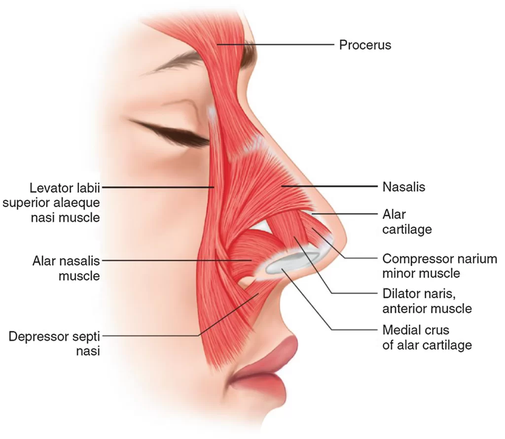
The Compressor Naris muscle is part of the nasalis muscle. The transverse part (compressor naris) arises from the maxilla, above and lateral to the incisive fossa; its fibers proceed upward and medially, expanding into a thin aponeurosis which is continuous on the bridge of the nose with that of the muscle of the opposite side, and with the aponeurosis of the Procerus.
Table of Contents
Origin:
The nasalis muscle is comprised of two regions: the transverse and the alar. The transverse section arises from the maxilla and covers the bridge of the nose. The alar is attached to the greater alar (nostril) cartilage, which is situated between the tissue of the nostril and the lateral cartilage.
Insertion:
The transverse part (compressor naris) arises from the maxilla, above and lateral to the incisive fossa; its fibers proceed upward and medially, expanding into a thin aponeurosis which is continuous on the bridge of the nose with that of the muscle of the opposite side, and with the aponeurosis of the Procerus. It compresses the nostrils and may completely close them.
Nerve supply:
Nasalis muscle is innervated by the facial nerve (7th cranial nerve).
Blood supply:
Arterial blood is supplied to the compressor naris via branches of the facial artery and the infraorbital branch of the maxillary artery.
Muscle action:

The nasalis is a sphincter-like muscle of the nose whose function is to compress the nasal cartilage. It is the muscle responsible for “flaring” the nostrils. Some people can use it to close the nostrils to prevent the entry of water when underwater.
Clinical Importance
Cleft lip and cleft palate
One of the main muscles that are incorrectly created or inserted with cleft lip and cleft palate deformity is the nasalis muscle.
During reconstructive surgery, the head of the transverse section must be located so that it may be surgically sutured (joined to) the nasal septum.
For improved symmetry, the origin at the maxilla may also be moved.
Facial nerve testing
The nasalis muscle can be utilized to assess the facial nerve because it is superficial. It may be used to evaluate the zygomatic branches specifically.
In Bell’s palsy compressor naris muscle is commonly paralysis.
Variations
The amount of the nose’s muscles varies from person to person and might even be completely nonexistent. The amount of the nose’s muscles varies from person to person and might even be completely nonexistent.
FAQs
The nasalis muscle, often referred to as the compressor naris, is situated inside the nose. It functions as a muscle with a ring-shaped sphincter. The muscle compresses nasal cartilage, as suggested by its other name. Additionally, it raises the corners of the nostrils while depressing the nose’s tip.
The lower surface of the nose’s nasal cartilages are covered by the nasalis muscle. The compressor naris muscle, which makes up the transverse portion, originates from the maxilla, above and lateral to the incisive fossa.
The zygomatic and temporal branches of the facial nerve (CN VII) supply the compressor narium minor muscle with its nerve fibres.