Dorsal Interossei muscle
Table of Contents
Dorsal Interossei Muscle Anatomy
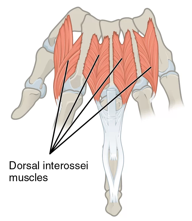
In human anatomy, the dorsal interossei (DI) are four muscles in the back of the hand that act to abduct (spread) the index, middle, and ring fingers away from the hand’s midline (ray of middle finger) and assist in flexion at the metacarpophalangeal joints and extension at the interphalangeal joints of the index, middle and ring fingers.
Origin
The origins are as follows,
- 1st DI originates from the adjacent sides of the 1st and 2nd metacarpals.
- 2nd DI originates from the adjacent sides of the 2nd and 3rd metacarpals.
- 3rd DI originates from the adjacent sides of the 3rd and 4th metacarpals.
- 4th DI originates from the adjacent sides of the 4th and 5th metacarpals.
Insertion:
It inserts on the proximal phalanges and extensor expansions.
Nerve supply:
The Deep branch of the ulnar nerve supplies the muscle.
Blood supply
The Dorsal and Palmar metacarpal artery supplies the muscle.
Action
It abducts fingers from the center of the third digiti. Both, palmar and dorsal interossei flex the metacarpophalangeal joints and extend the interphalangeal joints.
Function
The main function of the dorsal interossei is to abduct the fingers 2-4 in a longitudinal axis, moving them away from each other. Put simply, these muscles move the fingers in the following way:
The dorsal interossei 1-2 pull the digits 2-3 laterally (radial abduction),
The dorsal interossei 3-4 move the digits 3-4 medially (ulnar abduction).
The 3rd digit can be abducted both medially and laterally depending on which dorsal interosseous muscle contracts.
In addition to their main function, the dorsal interossei contribute to the flexion in the metacarpophalangeal (MCP) joints and extension in the proximal (PIP) and distal interphalangeal (DIP) joints. Compared to other muscles of the hand acting on these joints, the contribution of dorsal interossei here is rather negligible, but still important in terms of stabilization of these joints.
Strengthening exercise
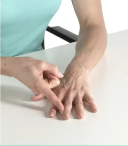
Isometric Finger Abduction Exercise:
- Sit upright in a chair.
- Place your affected arm on a table with your palm facing down and your fingers flat.
- Place the index finger from your unaffected hand on the thumb side of your middle finger.
- Try to move your middle finger towards your thumb, using your opposite hand to resist the movement.
- Keep your palm and forearm flat on the table throughout the exercise.
- Repeat the same on the other fingers.
Stretching exercise:

The dorsal interossei manus (of the central compartment group) are stretched with adduction of the index and ring fingers and with ulnar and radial abduction of the middle finger at the metacarpophalangeal joints.
Examination of the Muscle:
Examination of the muscle can be done while doing the action of the muscle. This muscle acts as an abductor of the fingers. These muscles can be resisted manually or by using a rubber band to check the strength of the muscle.
Related pathology
Exercise-induced chronic compartment syndrome in the first dorsal compartment is an uncommon entity and relatively rare condition that is not very well understood. It is a usually activity-related condition and is associated with decreased function of muscle with intracompartmental swelling. We present a case with proven exercise-induced raised compartment pressure that responded well to surgical fasciotomy.

