
Hip Joint
Table of Contents
Introduction
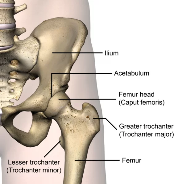
~The hip joint (see the image below) is a ball-and-socket type of synovial joint.
~The ball is the femoral head, and the socket is the acetabulum. The hip joint is the articulation of the pelvis with the femur, which connects the axial skeleton with the lower extremity.
~The hip joint is a ball and socket synovial joint, formed by an articulation between the pelvic acetabulum and the head of the femur.
Articulating Surfaces
~The head of the femur articulates with the acetabulum of the hip bone to form the hip joint.
~the head of the femur forms more than half a sphere,and is covered with the hyline cartilage except at the fovea capitis.
~the acetabulum presents a horseshoe-shaped, lunate articular surface, an acetabular notch, and an acetabular fossa.
~the lunate surface is covered with cartilage. Though the articular surfaces on the head of the femur and on the acetabulum are reciprocally curved, they are not co-extensive.
Stability
~The hip joint is unique in having a high degree of stability as well as mobility.the stability or strength depends upon:-
- Depth of the acetabulum and the narrowing of its mouth by the acetabular labrum.
2. Tension and strength of ligaments.
3. Strength of the surrounding muscles.
4. Length and obliquity of the neck of the femur.
5. atmospheric pressure
Ligaments:-
- The fibrous capsule
2. The iliofemoral ligament
3.The pubofemoral ligament
4.The ischiofemoral ligament
5. The ligament of the head of the femur
6.The acetabular labrum
7. The transverse acetabular ligament
Fibrous Capsule:
~it is attached on the hip bone to the acetabular labrum including the transverse acetabular ligament, and to bone above and behind the acetabulum, and on the femur to the intertrochanteric line in front, and 1 cm medial to the intertrochanteric crest behind.
~Anterosuperiorly:
~The capsule is thick and firmly attached. this part is subjected to maximum tension in the standing posture.
~posteroinferiorly:
~the capsule is thin and loosely attached to the bone.
~The capsule is made up of two types of fibers.
~the outer fibers= longitudinal
the inner are =circular called as Zona orbicularis.
~the joint cavity communicates with a bursae lying deep to the tendon of psoas major.
*Iliofemoral ligament:-
~it is inerverted Y-shaped ligament of bigelow, lies anteriorly.
~it is one of the strongest ligament of the body.
~it prevents the trunk for falling backward in the standing posture.
~it is triangular in shape.
Pubofemoral ligament:
~it is supports the joint inferomedially.
~it is also triangular in shape.
~superiorly,it is attached to the iliopubic eminence,the obturator crest and the obturator membrane.
~inferiorly,it merges with the anteroinferior part of the capsule and with the band of the iliofemoral ligament.
Ischiofermoral ligament:
~it covers the joint posteriorly.
~it is fibers or twisted and extend from the ischium to the acetabulum.
~the fibers of the ligament form the zona orbicularis.
~some of them are attached to the greater trochanter.

*ligament of the head of the femur:-
~it is round ligament or ligamentum teres is a flat and triangular ligament.
~the apex is attached to the fovea capitis,and the base to the transverse ligament and the margins of the acetabular notch.
*Acetabular notch:-
~it is a fibrocartilaginous rim attached to the margings of the acetabulum.
~it narrows the mouth of the acetabulum.
~this helps in holding the head of the femur in position.

Transverse ligament:
~it is a part of acetabular labrum which bridges acetabular notch.
~the notch is thus converted into a foramen which transmits acetabular vessels and nerves to the joint.
Blood supply:
1.Obturator artery
2.medial circumflex artery
3.lateral cicumflex artery
4.two gluteal arteries


Nerve supply:
- Femoral nerve
2. obturator nerve
3.Superior gluteal nerve

Muscles and Movement:-
- Hip Flexion: Psoas major and Iliacus muscle , Pectineus muscle, Rectus femoris muscle, sartorius muscle.
- Hip Extension: Gluteal maximus muscle and Hamstring Muscle
- Adduction: Adductor Muscle group – Adductor longus, Brevis and magnus other supporting muscles are Pectineus and Gracilis.
- Abduction: Gluteal medius and Gluteal Minimus muscle, Iliotibial band and sartorius muscle.
- Internal Rotation: Iliotibial Band and anterior fibres of the Gluteal medius and minimus
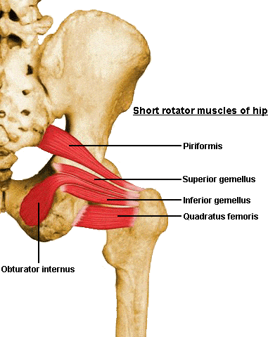
- External rotation: Two obturators, two gemelli & the quadratus femoris, piriformis, Gluteal maximus and sartorius


18 Comments