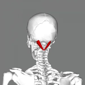
Plantaris Muscle
Table of Contents
Plantaris Muscle Anatomy
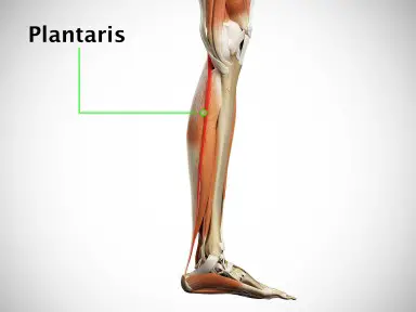
one of the superficial muscles of the superficial posterior compartment of the leg, one of the fascial compartments of the leg.
Origin
Lateral supracondylar ridge of femur above lateral head of the gastrocnemius muscle.
Insertion
endo calcaneus (medial side, deep to gastrocnemius tendon) .
Nerve supply
Plantaris is innervated by the tibial nerve, which is a branch of the sciatic nerve. The tibial nerve arises from the S1 and S2 spinal nerves.
Blood supply
Plantaris receives blood from the lateral sural artery, a branch of the popliteal artery. Its deep surface is supplied by the superior lateral genicular artery from the popliteal artery. The distal part of the plantaris tendon receives blood from the calcaneal branches of the posterior tibial artery.
Actions


Plantar flexes foot and flexes the knee.
Strengthening exercise
Single-Leg Heel Raise
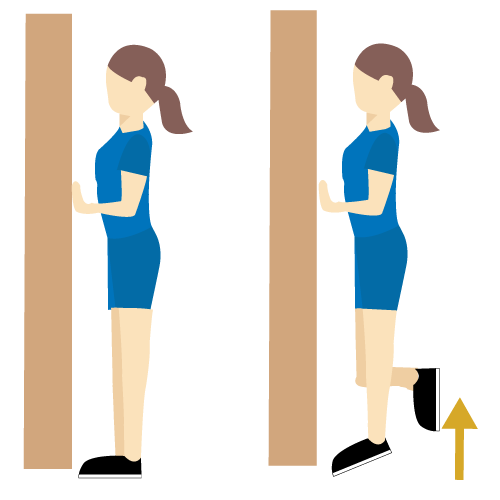
Stand barefoot on the balls of your feet with your heels hanging off a step. Hold on to a wall or doorframe for balance if necessary, but don’t use your hands for upward assistance. Lift one leg off the ground, and perform single-leg heel raises, also known as calf lifts, with the other. Move through a complete range of motion, from as low as you can go to as high as you can go. Try to do as many as you can with a full range of motion. Repeat on the other leg.
Weight-Bearing Lunge Test
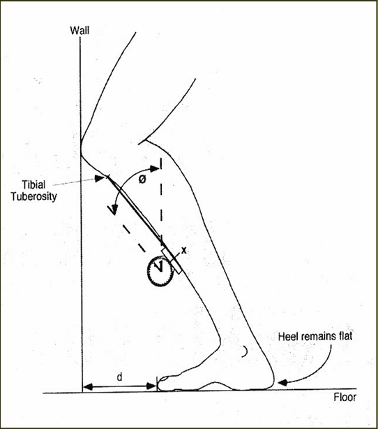
With your toes facing a wall, place one foot roughly a hand width away. Keeping your heel flat on the ground, bend your knee as if you were lunging into the wall. If your knee cannot touch the wall without your heel lifting, move it closer and try again. If your knee easily touches the wall, move your foot back and repeat. The idea is to find the distance where your knee can just barely touch the wall without your heel lifting. This is your dorsiflexion range.
Palpation
The popliteal fossa and the medial aspect of the triceps surae group’s common tendon can both be palpated to feel the muscle belly. The practitioner applies pressure to the plantar aspect of the foot with the forearm while covering the heel of the prone patient with their leg bent to roughly 90 degrees.
This allows for simultaneous resistance to plantarflexion of the foot and flexion of the knee. The muscle may be felt superior to and medial to the gastrocnemius muscle’s lateral head in the popliteal fossa. Its tendinous part may be felt all the way from the Achilles tendon’s medial side to the calcaneal insertion.
Clinical Importance
Injury
Injuries to the plantaris muscle and tendon, also known as “tennis leg,” have generated debate in the literature despite their small size.
A very common clinical issue is tennis leg. It has previously been linked to a number of causes, such as plantaris tears, soleus tears, medial head of gastrocnemius tears, or a mix of them. Running and jumping are the most common ways that the injury happens, and it generally happens as a result of an eccentric load applied to the ankle when the knee is extended.
Subjectively, the patient may describe direct trauma to the calf region, even when the injury is the consequence of an indirect mechanism. Frequently, the athlete feels like they got hit on the calf by an item, such as a ball or piece of equipment.
Calf soreness may be so severe that it prevents the athlete from continuing to play or it may just last for the duration of the exercise. Usually, the next day or after resting, this ache gets worse. Swelling that may reach the foot and ankle may be present in addition to the discomfort. Prolonged plantarflexion and any attempt at active or passive dorsiflexion will cause severe pain.

