Scalene Posterior Muscle
Scalene Posterior Muscle Anatomy
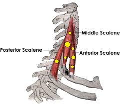
The scalene muscles are three paired muscles (anterior, middle, and posterior), located in the lateral aspect of the neck to the floor of the posterior triangle of the neck.
a group of three pairs of muscles in the lateral neck, namely the anterior scalene, middle scalene, and posterior scalene.
The posterior tubercles of the transverse processes of the final three or four cervical vertebrae are the origin of the posterior scalene muscle. It inserts on the second rib’s anterior face.
Origin
transverse processes of C4 – C6.
Insertion
2nd rib.
Nerve supply
The anterior branches of the cervical spinal nerves, which run from C3 to C8, innervate the scalene Posterior muscles.
C6, C7, C8.
Action

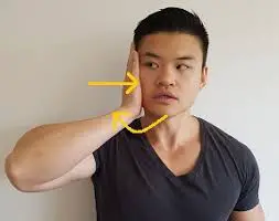
Elevate 2nd rib, and tilt the neck to the same side.
Anatomical Variation
Some of the fibres of the posterior scalene muscle can unite with the middle scalene or the first intercostal muscle to form a single muscular tissue that may involve the cervical vertebrae from C3 to C7. Finally, it may come into contact with the third thoracic rib. This muscle might not exist in certain individuals.
Brachial plexus branches like C7 and C8, which end on the posterior scalene muscle, have the potential to pierce the middle scalene. According to several writers, the posterior scalene muscle may consist of two layers: a dorsal and a ventral section.

