Temporoparietalis Muscle
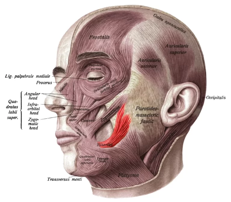
It is a distinct muscle located just above the auricularis superior. It is located inferior to the epicranial aponeurosis of the occipitofrontalis muscle.
The epicranius is a part of two belly muscles – The temporoparietalis and occipitofrontalis.
Table of Contents
Origin:
Auriculares muscles.
Insertion:
galea aponeurotica.
Nerve supply:
temporal branches of the facial nerve.
Action:
Fixes galeal aponeurosis .
It also helps to elevate the ear with auricularis superior muscle.

Clinical importance:
If Facial nerve damage where Bell’s palsy-related condition temporoparietalis muscle paralysis.
This muscle may be used in reconstructive ear surgery.
FAQ
An aponeurosis shared by the auricularis and the temporalparietalis muscle on either side gives origin to the scalp muscle. It inserts into the galeal aponeurosis by passing superiorly. It acts to repair the aponeurosis of the galea. The facial nerve’s temporal branch is called the nerve to temporoparietalis.
The epicranial aponeurosis is drawn toward the front of the skull by the temporoparietalis. It is innervated by the facial nerve, Cranial Nerve VII, through its temporal branches. The posterior auricular artery and the superficial temporal artery supply it.
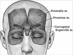
The forehead, top, and upper-rear portions of the skull are covered by the two parts of the epicranial muscle, commonly known as the epicranius. Forehead wrinkles are made possible by the frontalis region, which regulates eyebrow and forehead movement.
A strip of tendon called the galea, or galea aponeurotica, forms the epicranium above the skull’s dome. The frontalis and occipitalis are the two muscles that are linked to the galea from behind and in front. Here, on the occipital bone, above the superior nuchal line, is where the occipitalis muscle originates.

