Adductor Pollicis Muscle
Table of Contents
Adductor Pollicis Muscle Anatomy
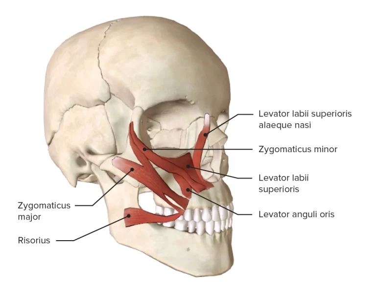
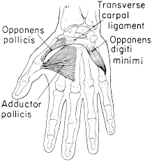
Adductor Pollicis is a fleshy, flat, triangular, and fan-shaped muscle deep in the thenar compartment beneath the long flexor tendons and the lumbrical muscles at the center of the palm.
It is situated in the palm and plays a vital role in the movement of the thumb. The term “pollicis” refers to the thumb, and “adductor” signifies its action of adduction, which is bringing the thumb towards the midline of the hand.
Origin:
It originates from 2 heads,
Transverse head: arises from the anterior body of the third metacarpal
Oblique head: arises from the bases of the second and the third metacarpals and the adjacent trapezoid and capitate bones.
Insertion:
It inserts on the medial side of the base of the proximal phalanx of the thumb and the ulnar sesamoid.
Nerve supply:
The adductor pollicis muscle is innervated by the deep branch of the ulnar nerve, specifically the deep motor branch.
Blood supply:
The deep palmar arch supplies the muscle.
Action:
It adductions the thumb at the carpometacarpal joint.
Function:
The adductor pollicis is the most powerful of the intrinsic muscles of the hand. Its main function is the adduction of the thumb which is the movement of the thumb towards the index finger from an abducted position.
This action is essential for functions that require pinching and gripping. Additionally, the adductor pollicis aids the later stages of the opposition of the thumb.
Relations
The thenar muscles are shallower and smaller than the adductor pollicis muscle. The flexor tendons of the index finger, the flexor pollicis brevis muscle, and the first lumbrical muscle cross the adductor pollicis on its ventral surface. The muscle is connected to, or occasionally even united with, the first dorsal interosseous muscle on its dorsal face. A route that is used by the radial artery, deep palmar arch, and deep branch of the ulnar nerve is also formed by the two heads of the adductor pollicis.
Exercise of Adductor Pollicis Muscle
Adductor Pollicis Muscle exercises mainly stretching and strengthening exercises.
Strengthening exercise
Strengthening the adductor pollicis muscle can be achieved through targeted exercises. Here’s an exercise that specifically targets the adductor pollicis muscle:
Thumb Opposition Exercise:
Begin by placing your hand on a flat surface, such as a table, with your palm facing downward.
Start with your thumb and pinky finger spread apart.
Slowly bring your thumb across your palm toward your pinky finger, attempting to touch the tip of your thumb to the tip of your pinky finger.
Hold this position for a few seconds, maintaining the contact between your thumb and pinky.
Slowly return your thumb to the starting position, away from your pinky finger.
Repeat this movement for a designated number of repetitions.
Perform the exercise on both hands.
This exercise specifically targets the adductor pollicis muscle by emphasizing thumb adduction and opposition. It helps to strengthen the muscle and improve its function.
Stretching exercise:
The adductor pollicis (of the central compartment group) is stretched with abduction and extension of the thumb at the metacarpophalangeal joint.
Clinical Significance
In some clinical conditions, such as nerve injuries or muscular disorders, the adductor pollicis muscle may be affected, leading to weakness or loss of function in thumb adduction. This can impact the hand’s overall dexterity and fine motor skills.
Physical therapists and hand specialists may prescribe specific exercises to strengthen and rehabilitate the adductor pollicis muscle, depending on the individual’s condition.
Deformity:
Upper limb deformity in cerebral palsy is a consequence of an imbalance between spastic and paretic muscles.
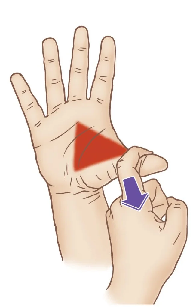
Froment’s sign.
Variations
There are nine distinct fascicles that may be seen inside the two heads of the adductor pollicis. There is significant variety in the precise origins and insertions of the nine distinct fascicles.
The muscle consists of two layers of fascicles in each head. There are two palmar and two dorsal fascicles in the transverse head and three dorsal and two palmar fascicles in the oblique head. The biomechanical characteristics of each thumb vary due to a variety of small, reportable variations among the nine distinct fascicles.
Embryology
The upper limb develops as a limb bud of mesodermal tissue from somites and the lateral plate starting on day 26 of week 4 of the foetal period. The limb develops from proximally to distally and is regulated by many signalling centres that generate distinct variables that govern differentiation. The zone of polarising activity (ZPA) controls radioulnar limb formation by producing sonic hedgehog protein (SHH), the apical ectodermal ridge (AER) controls proximal-distal signalling by inducing differentiation of underlying mesoderm and programmed cell death of interdigital tissue, and the Wnt pathway controls the ventral/dorsal limb axis. Developmental abnormalities pertaining to the upper limb may result from disturbances to any one of these three processes.
The full construction of hand structures starts at day 36, when chondrogenic forms of the digits appear. Early muscular masses start to develop across the hand and upper extremities during the sixth and seventh week. At this point, the development of the adductor pollicis and other intrinsic hand muscles starts. Week 7 sees the onset of interdigital apoptosis, which permits the development of distinct digits. Week 8 sees the ossification of the bones in the upper extremities. Developmental anomalies of the upper limb are likely to occur between weeks 3 through 8, as most upper limb development is finished by that time.
FAQ
The adductor pollicis’ primary job is to adduct the thumb. Adduction in this context refers to moving the thumb into an opposing position at the palm’s centre due to the thumb’s modified plane in regard to the palm and other digits.
The hand’s triangle intrinsic muscle is called the adductor pollicis. Along with the abductor pollicis, flexor pollicis brevis, and opponens pollicis, it is a member of the thenar muscles group.
The adductor pollicis muscle is tested for function using the Froment’s sign. If the adductor pollicis muscle is weak, the thumb IP joint will flex when the thumb and index finger are pinched together against resistance.
The hand’s intrinsic muscle is the adductor pollicis. It has two heads and is shaped like a triangle. The deep palmar arch is formed by the radial artery passing anteriorly through the area between the two heads.
To palpate the adductor pollicis, locate the muscle on the palm side of the hand near the base of the thumb. Place your thumb on the palm side of the first metacarpal bone and your index finger on the dorsal side. Squeeze gently, feeling for the muscle contraction beneath your fingers.