Buccinator Muscle
Table of Contents
Buccinator Muscle Anatomy
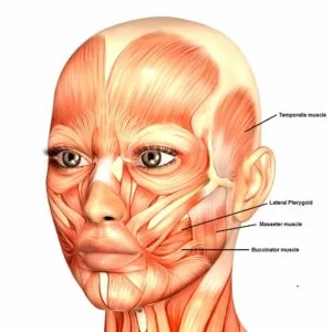
The buccinator muscle is a thin, four-sided facial muscle that is located in each of the cheeks. The buccinator muscle is a facial muscle located in the cheeks. The main action of this muscle is to compress the cheek against the teeth.
Origin
The upper fiber originates from maxilla, opposite molar teeth.
The lower fiber originates from mandible, opposite molar teeth.
The middle fiber originates from pterygomendibular raphe.
Insertion
The upper fiber inserts straight to the upper lip.
The lower fiber inserts straight to the lower lip.
The middle fiber decussate before passing to the lips.
Nerve supply
The buccal branch of the facial nerve (cranial nerve VII). Sensory innervation is supplied by the buccal branch (one of the muscular branches) of the mandibular part of the trigeminal (cranial nerve V).
Blood supply
Buccinator receives arterial blood supply mainly from the buccal artery, a branch of the maxillary artery; and some branches of the facial artery.
Structure
It arises from the outer surfaces of the alveolar processes of the maxilla and mandible, corresponding to the three pairs of molar teeth and in the mandible, it is also attached upon the buccinator crest posterior to the third molar;and behind, from the anterior border of the pterygomandibular raphe which separates it from the constrictor pharyngis superior.
The fibers converge toward the angle of the mouth, where the central fibers intersect each other, those from below being continuous with the upper segment of the orbicularis oris, and those from above with the lower segment; the upper and lower fibers continue forward into the corresponding lip without decussation.
Innervation
Motor innervation is from the buccal branch of the facial nerve (cranial nerve VII). Sensory innervation is supplied by the buccal branch (one of the muscular branches) of the mandibular part of the trigeminal (cranial nerve V).
Function
Its purpose is to pull back the angle of the mouth and flatten the cheek area, which aids in holding the cheek to the teeth during chewing. This action causes the muscle to keep food pushed back on the occlusal surface of the posterior teeth, as when a person chews. By keeping the food in the correct position when chewing, the buccinator assists the muscles of mastication.
It aids whistling and smiling, and in neonates it is used to suckle.
Action

flattens cheek against gums and teeth and prevents accumulation of food in the vestibule.
Strengthening exercise
1 Place your fingers over each cheekbone.
2 Gently lift the skin until taut.
3 Open your mouth to form an elongated “O”; you should feel resistance in your cheek muscles.
4 Hold for 5 seconds.
5 Complete 10-15 sets.
Related pathology
In Bell’s palsy buccinator muscle commonly paralysis.
FAQs
When eating, the buccinator keeps the cheeks compressed against the teeth while maintaining their tightness. Additionally, it helps the tongue keep the food bolus in the middle of the mouth cavity.
The buccinator mechanism, which consists of the orbicularis oris muscle, the buccinator, and the pharyngeal constrictor, plays a crucial part in the orofacial functions of swallowing, sucking, whistling, chewing, vowel pronouncing, and kissing.
The main facial muscle that supports the cheek is called the buccinator. It helps in chewing by holding the cheek against the teeth.
Early electromyographic (EMG) assessment of the buccinator in humans revealed that it was consistently but irregularly active during the majority of oral processes, such as sucking, blowing, swallowing, smiling, and speaking, typically together with the orbicularis oris muscle.

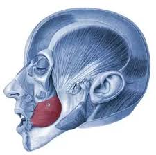
Great first picture. Never seen more beautiful skinned human face! Great drawer you have!!!
And the emphasizing of the muscle with green is so helpful while learning!!! Thank you!