The intrinsic muscles of the hand
Table of Contents
Introduction
The intrinsic muscles of the hand serve the function of adjusting the hand during gripping and also for carrying out fine skilled movements. The origin and insertion of intrinsic muscles are within the territory of the hand.
There are 20 muscles in the hand. These are:
- Four thenar muscles
Abductor pollicis brevis
Flexor pollicis brevis
Opponents pollicis
Adductor pollicis - Four hypothenar muscles
Palmaris brevis
Abductor digiti minimi
Flexor digiti minimi
Opponents digiti minimi - Four lumbricals
- Four palmar interossei
- Four dorsal interossel
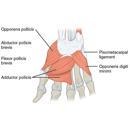
Four thenar muscles
Abductor pollicis brevis
Origin
The Abductor pollicis brevis muscle originates from the Tubercle of the scaphoid, Tubercle of the trapezium, flexor retinaculum
Insertion
The Abductor pollicis brevis muscle is inserted on the Lateral side of the base of the proximal phalanx of the thumb and Some fibers are inserted into the dorsal digital expansion.
Nerve supply
The nerve supply of the Abductor pollicis brevis muscle is the Median nerve(C8, T1)
Blood supply
The blood supply of the Abductor pollicis brevis muscle is the Superficial palmar branch of the radial artery
Action
Abduction of thumb at the metacarpophalangeal joints and carpometacarpal joints. Abduction is associated with medial rotation.
Flexor pollicis brevis
Origin
The Flexor pollicis brevis muscle originates from the Flexor retinaculum, trapezoid, and capitate bones
Insertion
The Flexor pollicis brevis muscle is inserted on the Base of the proximal phalanx of the thumb
Nerve supply
The nerve supply of the Flexor pollicis brevis muscle is the Median nerve
Blood supply
The blood supply of the Flexor pollicis brevis muscle is the superficial palmar branches of the radial artery.
Action
Flexion of the metacarpophalangeal joint of the thumb
Opponens pollicis
Origin
The origin of the Opponens pollicis muscle is the Flexor retinaculum
Insertion
The Opponens pollicis muscle is inserted on the Lateral half of the palmar surface of the shaft of the metacarpal bone of the thumb
Nerve supply
The nerve supply of the Opponens pollicis muscle is the Median nerve
Blood supply
The blood supply of the Opponens pollicis muscle is the Superficial palmar branch of the radial artery
Action
Pulls thumb medially and forward across palm (opposes thumb towards the fingers)
Adductor pollicis
Origin
The oblique head of the Adductor pollicis muscle originated from the Bases of 2nd-3rd metacarpals;
transverse head of the Adductor pollicis muscle originated from the Shaft of 3rd metacarpal
Insertion
The Adductor pollicis muscle is inserted on the Base of the proximal phalanx of the thumb on its medial aspect
Nerve supply
The nerve supply of the Adductor pollicis muscle is the deep branch of the ulnar nerve which ends in this muscle
Blood supply
The blood supply of the Adductor pollicis muscle is the deep palmar arterial arch, formed by the anastomosis of the radial artery and the deep palmar branch of the ulnar artery.
Action
Adduction of thumb
Four hypothenar muscles
Palmaris brevis
This muscle is superficial and lies just under the skin.
Origin
The origin of the Palmaris brevis muscle is the Flexor retinaculum and palmar aponeurosis.
Insertion
The Palmaris brevis muscle is inserted on the Skin along the medial border of the hand
Nerve supply
The nerve supply of the palmaris brevis muscle is the Superficial branch of the ulnar nerve(C8, T1)
Blood supply
The blood supply of the palmaris brevis muscle is the Superficial palmar arch
Action
It Helps in gripping by making the hypothenar eminence more prominent, and by Wrinkling the skin over it.
Abductor digiti minimi
Origin
The origin of the Abductor digiti minimi muscle is the Pisiform bone
Insertion
The Abductor digiti minimi muscle inserted on the Ulnar side of the Base of the proximal phalanx of the little finger
Nerve supply
The nerve supply of the Abductor digiti minimi muscle is the deep branch of the ulnar nerve
Blood supply
The blood supply of the Abductor digiti minimi muscle is the Ulnar artery
Action
Abduction at the metacarpophalangeal joint of the little finger.
Flexor digiti minimi
Origin
The Flexor digiti minimi muscle originates from the Hook of hamate bone and the Flexor retinaculum
Insertion
The Flexor digiti minimi muscle is inserted on the Ulnar side of the Base of the proximal phalanx of the little finger
Nerve supply
The nerve supply of the v muscle is the deep branch of the ulnar nerve
Blood supply
The blood supply of the Flexor digiti minimi muscle is the Ulnar artery
Action
At the little finger metacarpophalangeal joint Flexion.
Opponens digiti minimi
Origin
Thye Opponens digiti minimi muscle originates from the Hook of hamate bone and Flexor retinaculum
Insertion
The Opponens digiti minimi muscle is inserted on the Medial surface of the shaft of the fifth metacarpal bone.
Nerve supply
The nerve supply of the Opponens digiti minimi muscle is the deep branch of the ulnar nerve
Blood supply
The Blood supply of the Opponens digiti minimi muscle is by the deep palmar branch of the ulnar artery and deep palmar arch, which is the terminal branch of the radial artery
Action
Flexion of the fifth metacarpal and rotates it laterally(as in making the palm hollow).
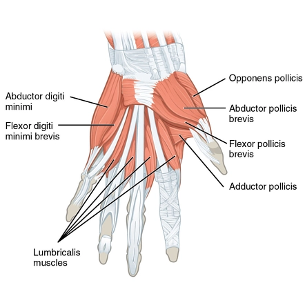
Lumbricals muscles
Lumbricals muscles are four small muscles that take origin from the tendons of the flexor digitorum profundus. They are numbered from the lateral side to the medial side.
Origin
The first lumbrical originates from the radial side of the tendon of the flexor digitorum profundus of the 2nd digit.
The second lumbrical originates from the radial side of the tendon of flexor dlgitorum profundus of 3rd digit
The third lumbrical originates from the Adjacent sides of the tendon of flexor dlgitorum profundus of 3rd and 4th digits
The fourth lumbrical originates from the Adjacent sides of tendons of flexor dlgitorum profundus 4th and 5th digits
Insertion
The first, second, third, and fourth lumbrical muscles tend backward on the radial side of the second, third, fourth, and fifth metacarpophalangeal joints respectively. They are inserted into the dorsal digital expansions of the corresponding digits.
Nerve supply
The nerve supply of the First and second lumbricals is the median nerve, and The third and fourth lumbricals are the deep branch of the ulnar nerve
Blood supply
The blood supply of the lumbrical muscles from the anastomotic network on the dorsal surface of the hand is known as the dorsal carpal arch. Its branches, first and second dorsal metacarpal arteries and dorsal digital arteries, supply the first and second lumbrical muscle.
The third and fourth dorsal digital arteries and second and third common palmar digital arteries (branches of the superficial palmar arch) supply the third and fourth lumbrical muscle.
Action
The lumbricals muscle Flexes metacarpophalangeal joints and extends the interphalangeal joints of the 2nd-5th digits
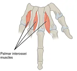
Palmar interossei
The Palmar interossei muscles are four small muscles placed between the shafts of the metacarpal bones. They are numbered from the lateral side to the medial side.
Origin
The First Palmar interossei muscle originates from the Medial side of the base of 1st metacarpal bone
The second Palmar interossei muscle originates from the medial half of the palmar aspect of the shaft of the 2nd metacarpal bone
The third Palmar interossei muscle originates from the Lateral part of the palmar aspect of the shaft of the 4th metacarpal bone
The fourth Palmar interossei muscle originates from the Lateral part of the palmar aspect of the shaft of the 5th metacarpal bone
Insertion
Each muscle of the Palmar interossei is inserted into the dorsal digital expansion of one digit. it may be attached to the base of the proximal phalanx of the same digit. The digits into which individual palmar interossei muscles are inserted are as follows.
First muscles: Medial side of the thumb
Second muscles: Medial side of the index finger
Third muscles: Lateral side of the fourth digit
Fourth muscles: Lateral side of the fifth digit
Nerve supply
The nerve supply of the palmar interossei is the Deep branch of the ulnar nerve(C8, T1)
Blood supply
Palmar interossei muscles receive arterial blood supply from the palmar metacarpal arteries and drain into the palmar metacarpal veins. The palmar metacarpal arteries derive from the deep palmar arch, which is comprised of the terminal part of the radial artery and the deep branch of the ulnar artery.
Action
Palmar interossei adduct fingers towards the center of the third digit or middle finger. In addition, they flex the digit at the metacarpophalangeal joint and extend the digit at the interphalangeal joints.
Dorsal interossei
Like the palmar interossei, the dorsal interossei are four small muscles placed between the metacarpal bones and are numbered from the lateral side to the medial side.
Origin
First dorsal interossei originate from the shafts of 1st and 2nd metacarpals bone
Second dorsal interossei originate from the shafts of 2nd and 3rd metacarpals bone
Third dorsal interossei originate from the shafts of 3rd and 4th metacarpals bone
Fourth dorsal interossei originate from the shafts of 4th and Sth metacarpals bone
Insertion
Each muscle of the dorsal interossei is inserted into the dorsal digital expansion of the digit, and into the base of the proximal phalanx of that digit. The digits into which individual muscles of the dorsal interossei are inserted are as follows.
First: Lateral side of the index finger
Second: Lateral side of the middle finger
Third: Medial side of, middle finger
Fourth: Medial side of the fourth digit
Nerve supply
The nerve supply of the dorsal interossei muscles is the Deep branch of the ulnar nerve(C8, T1)
Blood supply
The Nerve supply of the Dorsal Interossei muscles of the hand is the dorsal and palmar interossei artery
Action
Dorsal interossei abduct the fingers from the center of the third digit. dorsal interossei muscles flex the metacarpophalangeal joints and extend the interphalangeal joints
Clinical significance
Claw Hand
Claw Hand deformity is a condition in which the fingers are bent into a position that looks like a claw. It may affect all of the fingers or only some of them depending upon its cause. The fingers are usually bent or curved and the deformity may affect one hand or both hands. Claw Hand is a deformity with hyperextension of the metacarpophalangeal (MCP) joints and flexion of the interphalangeal (IP) joints due to weakness of the intrinsic muscles of the hand.
Types
Claw Hand deformity can be:
Complete claw hand deformity when it involves all the digits and therefore results from both Ulnar and Median Nerve Palsy.
Incomplete or Partial claw hand deformity that involves only the ulnar 2 digits and is referred to as an isolated Ulnar Nerve Palsy.
Ape Hand
The ape hand is a deformity in humans causing an inability to abduct or oppose the thumb thereby causing the thumb little or no abduction and opposition.
Exercise of the intrinsic muscles of the hand
Flap
Bend the knuckles while keeping the fingers straight.
Hold this position for 3 seconds, then bring it back to a straight position.

Hook
Bend the top two joints of your digits, keeping your knuckles straight.
Hold this position for 3 seconds, then bring them straight again.
Tents
Starting with your hand flat, palm down on the table, bend up and the knuckles keeping your fingers straight, then lower again and repeat.
Spreading the fingers
With your hand straight, bring the fingers apart.
Pulling the fingers back in
Bring your fingers back together.
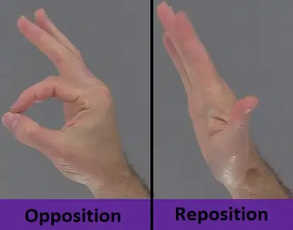
Opposition
Take your thumb and little finger towards each other.
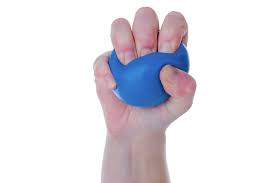
Ball Squeeze
Hold the squeeze ball in your hand and squeeze it into your hand until your fingertips reach your palm, then release and then squeeze again. repeat 20 times.
Thumb Pinch Strengthening
Use the putty, and roll it out into a ‘hot dog’ shape about 1-2 inches thick. using your thumb and index finger, pinch along the putty, making little indents along the length of the putty.
Do this 10 to 20 times.
Rubber Band Abduction
Place the rubber band over all four fingers. Do not include the thumb. Now spread your fingers apart as far as you can, hold for 5 seconds, then relax. Repeat 10 to 20 times.