
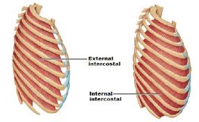
External intercostal muscles
External intercostal muscles are eleven in number on both sides.
origin:
Lower border of ribs.
Insertion :
Upper border of rib below.
Nerve:
intercostal nerves.
Actions:
Inhalation.
External intercostal muscles are eleven in number on both sides.
origin:
Lower border of ribs.
Insertion :
Upper border of rib below.
Nerve:
intercostal nerves.
Actions:
Inhalation.
Physiotherapist , Samarpan Physiotherapy Clinic, Vastral, Nirant Cross Road, Ahmedabad
Home Visit Treatment Also Available in Bapunagar Vastral Rabari Colony Char Rasta, CTM, Maninagar , Viratnagar , Nikol Nava Naroda And NearBy Area Of Ahmedabad.
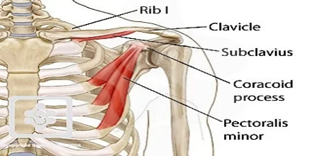
Subclavius Muscle Anatomy The subclavius is a small triangular muscle, placed between the clavicle and the first rib. Along with the pectoralis major and pectoralis minor muscles, the subclavius muscle makes up the anterior wall of the axilla. Origin The subclavius muscle originates from the first rib. Specifically, this muscle begins in the area between…
Levator labii superioris alaeque nasi Muscle Anatomy The levator labii superioris alaeque nasi muscle is translated from a Latin word that means “Lifter of both upper lips and wing of the nose “. It has the longest name of any muscle in the animal. Historically known as Otto’s muscle, it dilates the nostril and elevates…
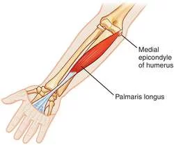
Palmaris longus Muscle Anatomy The palmaris longus is a muscle visible as a small tendon between the flexor carpi radialis and the flexor carpi ulnaris, although it is not always present. About 14% of people do not have it; however, communities of African, Asian, and Native American people may have different percentages. Grip strength is…
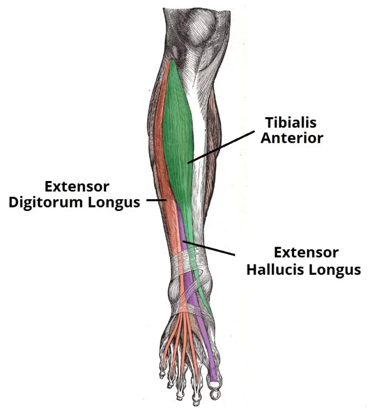
Tibialis anterior Muscle Anatomy Tibialis anterior is a muscle of the human body that is used for the movement of the ankle joint. It has its origin on the lateral surface of the tibia, and its insertion on the medial cuneiform and first metatarsal. The muscle is one of the superficial muscles of the sole…
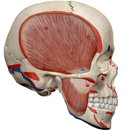
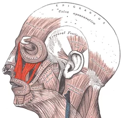
Introduction The muscles of mastication move your mandible during mastication and speech. The muscles of mastication are the Masseter, the Temporalis, The Lateral pterygoid, and the medial pterygoid. They develop from the mesoderm of the first branchial arch and are supplied by the mandibular nerve which is the nerve of the branchial arch. The muscles…

Teres Major Muscle Anatomy The teres major muscle is one of the six muscles within the scapulohumeral muscle group. This muscle is commonly confused as a rotator cuff muscle, but it is not because it does not attach to the capsule of the shoulder joint, unlike the teres minor muscle. Origin: It originates from the…