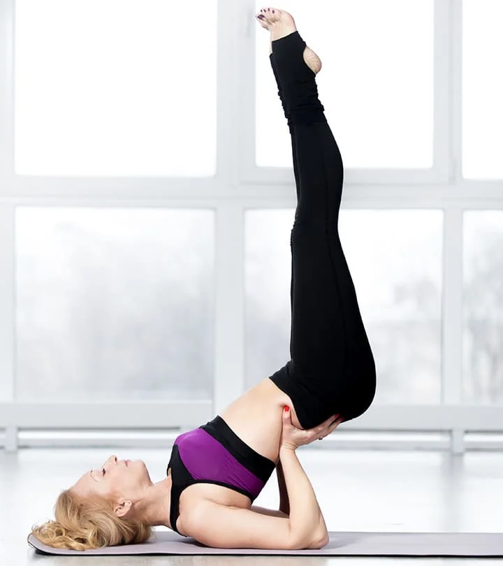
Muscle of medial compartment of the thigh
Table of Contents
Introduction
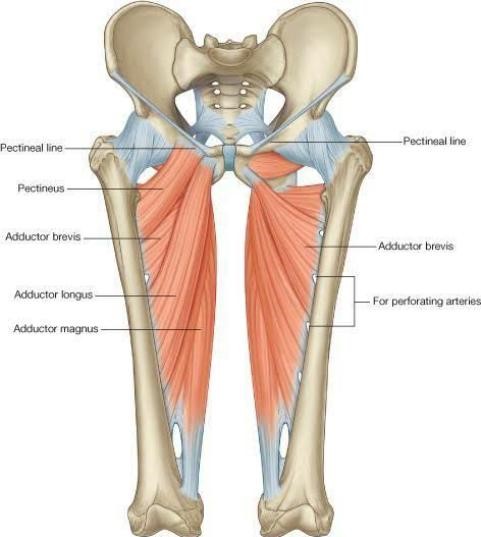
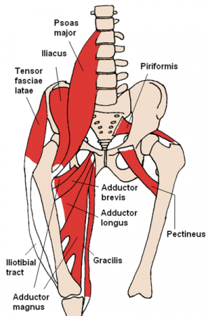
The hip adductors are a group of muscles located in the medial compartment of the thigh. These muscles include the adductor longus, adductor brevis, adductor Magnus, gracilis, and pectineus.
Due to their position, the hip adductors shape the surface anatomy of the medial side thigh. More specifically, these muscles extend from the anteroinferior external surface of the bony pelvis to the shaft of the femur and proximal part of the tibia.
The majority of hip adductor muscles are innervated by the obturator nerve (L2 to L4) and supplied by blood mainly via the branches of the femoral arteries and obturator arteries.
Adductor longus
The Adductor longus is a triangular muscle, forming the medial part of the floor of the femoral triangle. Adductor longus lies in the plane of the pectineus
Origin
Adductor longus originate from a narrow, flat tendon from the front of the body of the pubis at the angle between the pubic crest and the pubic symphysis. Sometimes the sesamoid bone is seen near its origin (rider’s bone)
Insertion
The linea aspera in the middle one-third of the shaft of the femur between the vastus medialis and the adductor brevis and adductor Magnus
Nerve supply
The nerve supply of the Adductor longus muscle is the Anterior division of the obturator nerve
Blood supply
The blood supply of the adductor longus comes from two arteries, the profunda femoris artery (a branch of the femoral artery) and the obturator artery (a branch of the internal iliac artery).
The proximal part of the Adductor longus muscle is supplied by the medial circumflex artery (branch of the profunda femoris artery).
Action
Powerful adductor of thigh at hip joint
The adductor muscles act as posture controllers
Adductor brevis
The Adductor brevis muscle lies behind the pectineus and the adductor longus muscle
Origin
a. Anterior surface of the body of the pubis
b. The outer surface of the inferior ramus of the pubis between the gracilis and obturator externus
muscle
c. Outer surface of the ramus of the ischium between gracilis and the adductor Magnus’s muscle
Insertion
The line extends from the lesser trochanter to the upper part of linea aspera. behind the upper part of the adductor longus muscle
Nerve supply
The nerve supply of The Adductor brevis muscle is the Anterior or posterior division of the obturator nerve
Blood supply
The blood supply of the adductor brevis muscle typically comes from the deep femoral artery (profunda femoris) and from its branch called the artery for the adductors. It can also be supplied partially from the medial circumflex femoral artery and obturator artery.
Action
Adductor longus, adductor brevis help in adduction and flexion of thigh at hip joint
Adductor Magnus
The Adductor Magnus is the largest muscle in this compartment. Because of its double nerve supply, it is also called a hybrid muscle
Origin
a. Inferolateral part of the ischial tuberosity
b. Ramus of the ischium
c. The lower part of the inferior ramus of the pubis
Insertion
a. Inferolateral part of the ischial tuberosity
b. Ramus of the ischium
c. Lower part of inferior ramus of the pubis
Nerve supply
Double nerve supply: Adductor part by posterior division of obturator nerve and
Hamstring part by tibial part of the sciatic nerve
Blood supply
The blood supply of the adductor Magnus’s muscles is the perforating branches of the deep femoral artery, passing through its oseo-aponeurotic openings.
Action
The Adductor part causes adduction of the thigh and the hamstring part helps in the extension of the hip and flexion of the knee joints
Gracilis
(Greek slender)
Origin
The medial margin of the lower half of the pubis body, the Inferior ramus of the pubis, and the Adjoining part of the ramus of the ischium
Insertion
The upper part of the medial surface of the tibia behind the sartorius and in front of the semitendinosus muscles
Nerve supply
The nerve supply of the Gracilis muscle is the Anterior division of the obturator nerve
Blood supply
The blood supply of the gracilis muscle is the medial circumflex femoral artery.
Action
Flexor and medial rotator of thigh at the hip joint. The Gracilis muscle is a weak adductor of the thigh.
Gracilis muscle is used for transplantation of any damaged muscles
Pectineus
The Pectineus is a flat, quadrilateral muscle It forms a part of the floor of the femoral triangle
Origin
The Pectineus muscle originates from the Pecten pubis, the Upper half of the pectineal surface of the superior ramus of the pubis, and the Fascia covering the pectineus
Insertion
The line is extended from the lesser trochanter to the linea aspera
Nerve supply
Double nerve supply: Anterior fibers by femoral nerve and Posterior fibers by anterior division of obturator nerve
Blood supply
the superficial part of the Pectineus muscle supplied by the medial circumflex femoral artery and the deep part of the Pectineus muscle supplied by the anterior branch of the obturator artery
Action
Flexor of thigh
Adductor of thigh
Clinical Importance
The adductor muscle strain ranks among the most common sports injuries (e.g. playing soccer, doing the splits, slipping on ice, etc.) and affects favorably the origin tendon in the pubic region.
It occurs by a disproportional strain of the muscles, often in combination with a poor warm-up and a lack of stretching. Hereby even ruptures and hemorrhages can occur.
Symptoms can include pain extending to the inguinal and knee region when stretching and straining the muscles. A sacroiliac joint mal-position can restrict the function of the hip adductors in the long run as well and should be addressed clinically.
The orthopedic pathologies mentioned above are to be distinguished from the neurogenic spasm of the adductor. This is a common symptom of spastic diplegia (Little’s disease). Because of the spasticity in the hip adductors, the affected children walk with adducted, flexed, and internally rotated legs (scissor gait).
Exercise for the muscle of the medial compartment of the thigh
Strengthening exercise
Wide stance squat
Set your feet a little bit wider than shoulder-width, turning your toes slightly outward.
Shift your weight back and slowly lower your hips until your thighs are parallel to the ground.
In a controlled motion, return to the starting position by pushing through the ground, feeling the contraction of muscles of your glutes and legs, including your adductors.
Repeat for 10 to 12 repetitions for 2 to 3 sets.

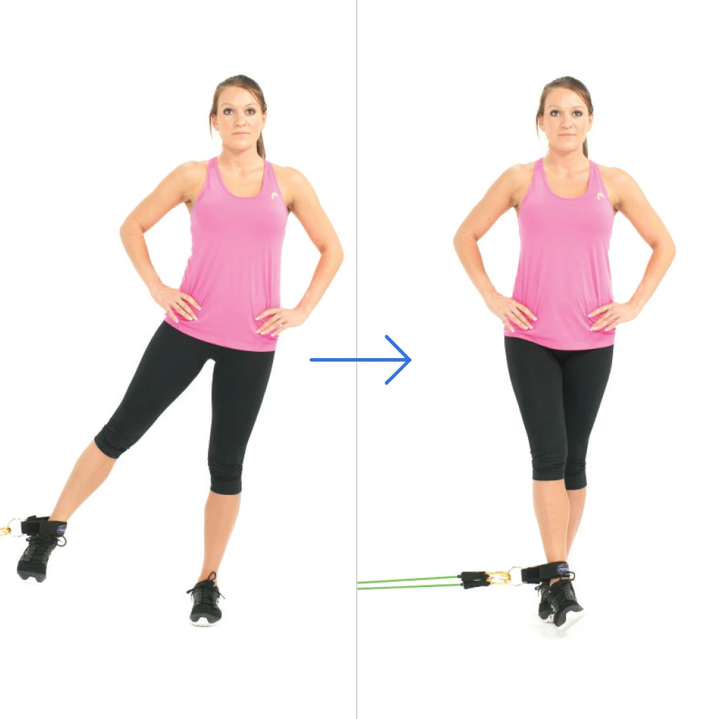
Standing banded adduction
Start by wrapping a resistance band around a solid anchor such as a power rack or another piece of equipment that is attached to the ground.
Standing with either side of your body facing the anchor point, wrap the band around your inner foot.
Adjust the tension of the resistance band by standing farther away from the anchor point or wrapping the resistance band more tightly.
Allow the resistance band to pull your leg to the side while resisting the movement.
To start the exercise, stand straight and bring your banded leg toward the center of your body, feeling a good contraction in your adductors.
Gently release your leg back to the side, with control.
Repeat for 10 to 12 repetitions for 2 to 3 sets.
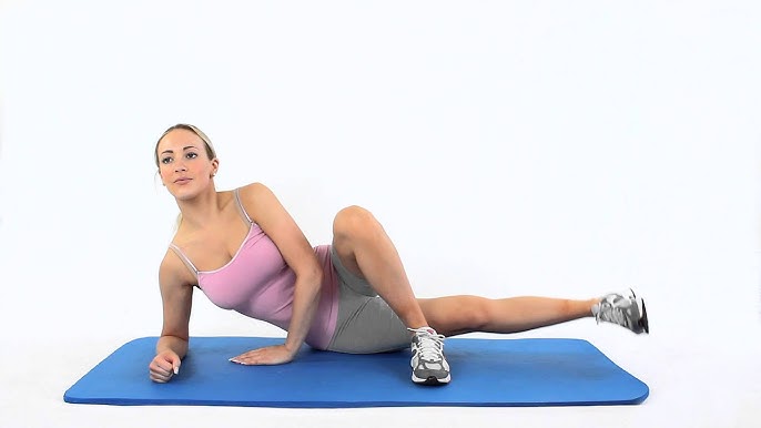
Side-lying adduction
Lie on your side on a mat. Ensure that your spine is in neutral and your hips are stacked. Keep your bottom arm folded under your head and your top hand on the ground in front of your stomach.
Bend your top knee so it points toward the ceiling and places the foot flat on the ground in front of your bottom leg.
Keeping your bottom leg long and foot flexed, lift your bottom leg off the ground, contracting your adductors.
Allow your leg to slowly return to the ground in a controlled motion.
Repeat for 10 to 12 repetitions for 2 to 3 sets.
Stretching exercise
Short Adductors can also be stretched in a sitting position.
Sit on the ground with your knees apart and the bottom of your feet together.
Keep your back straight and chest up.
Bend forward at the hips while accentuating your lumbar lordosis
Apply gentle pressure to your knees pushing them toward the ground.
hold this for the appropriate time and then relax.

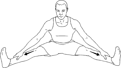
Long adductors can also be stretched in a sitting position.
This stretch also includes hamstrings biasing the medial hamstrings, semimembranosus, and semitendinosus muscles.
for both sides simultaneously stretch, sit with your legs straight out in front of you with your back straight.
Slowly work your legs apart as far as possible.
Hold this position and relax for a few seconds.
Now as you exhale bend forward at your hips until you can feel more resistance.
Be sure to keep your chest up and to maintain a lumbar lordosis
Hold this for the appropriate time.