Facial Nerve Palsy: Cause, Symptoms, Diagnosis, Treatment, Exercise
Table of Contents
What is facial nerve palsy?
- Facial nerve palsy is a term used to describe any kind of paralysis of facial muscles.
- Facial palsy is caused by damage to the facial nerve or cranial nerve 7 Which supplies the muscles of the face.
- It is categorized into two types based on the location of the casual pathology:
- Central facial nerve palsy :
- Occurs due to damage above the level of the facial nucleus.
- Peripheral facial nerve palsy :
- Occurs due to damage at the level or below the level of the facial nucleus.
Relevant Anatomical Course of Facial Nerve:
The facial nerve has a very complex course structure.
There are many branches of the facial nerve, which provide supply to sensory, motor, and parasympathetic fibers.
- so, basically, the course of the facial nerve is divided into 2 parts :
- Intracranial
- Extracranial
- it is divided according to the site from which the nerve will pass through it.
- Intracranial: Here, the course of the nerve will be through the cranial cavity and cranium itself.
- Extracranial: The course of the nerve will be through the face and neck, so by outside of the cranium.
- both the divisions are described below in detail.
Intracranial course :
- The facial nerve arises from the Pons, it is the area of the Brainstem.
- from here, it is divided into 2 roots :
- Motor and Sensory
- The motor root is large.
- The sensory root is small.
- both the roots travel through the internal acoustic meatus.
- internal acoustic meatus is a large or long opening of the Temporal bone.
- after this, both the roots will leave the internal acoustic meatus and will enter the Facial canal.
- into the facial canal, the following will occur :
- Both sensory and motor roots will fuse together and will form one single Nerve, which is called a Facial Nerve.
- next, The Facial Nerve will form the Geniculate Ganglion, which is the collection of the nerve cell bodies.
- so, after this The Facial nerve gives rise to the following :
- Greater Petrosal Nerve :
- it innervates the parasympathetic fibers of mucous glands and lacrimal glands.
- Nerve to Stapedius muscle :
- it innervates the motor fibers of the stapedius muscle which is situated in the middle ear.
- Chorda Tympani :
- it innervates the sensory fibers to the anterior 2/3 tongue.
- and also innervates parasympathetic fibers of submandibular and sublingual glands.
- after this, the Facial Nerve will exit the Facial canal via the Stylomastoid foramen.
- The stylomastoid foramen is located posterior to the styloid process of the temporal bone.
- so this is the intracranial course of the facial nerve.
Extracranial course:
- After exiting from the cranium or skull, the Facial nerve turns superiorly and reaches up to the anterior part of the outer ear.
- From here, the Facial Nerve will divide into some motor branches. this is described here.
- Posterior Aurical Nerve :
- It provides motor innervation to some muscles around the ear.
- some motor branches will also supply to the Digastric muscle mostly, posterior muscle belly.
- and some motor branches will also supply Stylohyoid muscle.
- Now, The Main Motor Root of the facial nerve will travel continuously anteriorly and inferiorly into the Parotid gland.
- Within the Parotid gland, the nerve terminates by diving into five main branches.
- The name of these five branches are as follow :
- Temporal Branch
- Zygomatic Branch
- Buccal Branch
- Marginal Mandibular Branch
- Cervical Branch
What are the types of Facial nerve palsy?
There are mainly 2 types:
Central facial nerve palsy
peripheral facial nerve palsy
Central Facial Nerve Palsy :
- Central facial palsy happens when some structures of the brain get damaged.
- The most common cause can be the condition of stroke.
- Here, The symptoms of facial palsy will be limited to the lower half of the face.
Peripheral Facial Nerve Palsy:
- In Peripheral facial palsy, there will be complete paralysis of one side of the face.
- so here, both the upper and lower half of the facial muscles will become affected.
What are the Possible Causes of Facial Nerve Palsy?
- Stroke
- Intracranial tumor
- Multiple sclerosis
- Syphilis infection
- HIV infection
- Hemorrhage
- Idiopathic cause
- Herpes Simplex viral infection
- compression by the tumor to the facial nerve can also cause nerve palsy.
- compression while surgical removal of a tumor
- Ramsay Hunt syndrome
- vestibulocochlear dysfunction
- Lyme disease
- Trauma – temporal bone fractures
- Otitis media
- Bell’s palsy
What is the possible pathology behind the condition?
- The facial nerve becomes inflamed or swallowed within the facial canal.
- There was limited space in the canal because it is a narrowed area.
- So the nerve gets compressed within it.
- And it occurs repeatedly, so the facial nerve losses its normal conductivity.
- And the muscles supplied by the nerve also lose their normal function.
Which are muscles get affected by Facial nerve palsy?
There is a number of facial muscles that may get affected by Facial nerve palsy, which is innervated by the facial nerve. So because of this motor and sensory, both functions will become affected.
it is described below in detail.
it depends on the type of facial palsy.
if you have central facial nerve palsy, your upper half of the facial muscles will remain normal.
but if you have peripheral facial nerve palsy, there will be affection on one side of facial muscles, so upper and lower half muscles both will be affected here.
here, facial muscles affected in peripheral facial palsy are described in detail.
Because of inflammation of the facial nerve, the motor and sensory functions will be affected.
- Affected motor functions :
- The main muscles get affected are as follows:
- Occipital frontalis:
- Action: Raises the eyebrows.
- Orbicularis oculi:
- Action: Closes the eyes.
- Corrugator and procerus :
- Action: Wrinkles skin between your Eyebrows and frowns.
- Zygomaticus major and minor, levatorangulioris ,levatorlabisuperioris :
- Action: Raises the corner of the mouth and upper lip.
- Buccinator :
- Action: keep the cheeks against the teeth during mastication and sucking Activities.
- Orbicularis Oris:
- Action: Closes the mouth.
- Risorius :
- Action: pulls the angle of the mouth backward, as seen in grinning.
- Nasalis :
- Action: Depresses the tip of the nose and helps in elevation of the corner of the nostrils.
- Depressor angulioris and Depressor labii inferioris :
- Action: pulls down the angle of the mouth and your lower lip.
- Mentalis :
- Action: wrinkles your chin.
- It is very important to muscles for drinking because it holds the lower lip on the cup or bottle to prevent dribbling.
- Affected sensory functions :
- The facial nerve also supplies the sensation of the pallet and anterior two / third of the tongue.
- Also supplies the salivary glands present in the mouth.
What kind of symptoms are seen in the patients diagnosed with Facial Palsy?
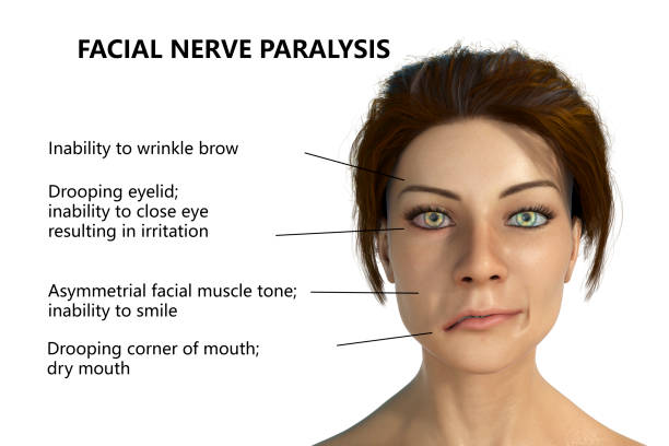
- Loss of facial expressions is seen.
- The dropping of the corner of the mouth and mouth sag are also seen.
- Creases are seen on the face and skin folds become smooth.
- The dropping of the eyebrow and wrinkles were also seen.
- The forehead is seen without furrowing.
- Closure of the eye becomes impossible or difficult for the patients.
- Bell’s phenomenon is seen.
- What is the Bell’s phenomenon?
- When the patient tries to close his eyes, there will be movement of the eyeball in the upward and slightly inward direction.
- It is called Bell’s phenomenon.
- Retraction of the mouth and pursuing of lips is not possible by the patient.
- Dribbling of saliva from the corner of the mouth is seen.
- Chewing and eating become difficult for the patients. because of paralysis of buccinator, there will be the accumulation of food into the mouth is seen.
- Sometimes speaking also become difficult for the patients.
- The patient feels heaviness and numbness in the face.
- Deviation of the tongue might be seen.
- In advance cases, loss of taste sensations in two/ third of the tongue is seen.
- Lacrimation might be seen reduced.
- Nasolabial folds become smooth and start deepening.
- So the loss of normal function of facial muscles is seen.
How to diagnose Facial Palsy?
- By asking about the previous and present history of the patient related to any viral infections.
- By assessing the functions of facial muscles during the examination.
- Or your doctor may suggest you for investigations like :
- Electromyography (EMG)
- This investigation will help to know about the functions of facial muscles.
- Strength Duration curves or SD curves and RD Test.
- Nerve conduction studies ( NCS):
- This investigation will help to know whether the stimulated muscles are Innervated, Denervated, or partially Denervated.
What is the Differential Diagnosis of Facial Palsy?
- Herpes zoster oticus :
- This disease also causes facial paralysis and hearing loss in the affected ear.
- Other Infections :
- For example Acute Otitis media, Lyme disease
- Multiple sclerosis :
- In this Disease, the immune system attacks the body’s nervous system.
- Gillian Barre syndrome :
- In this Disease, the immune system attacks the nervous system of the body.
- The condition may be triggered by bacterial or viral infection.
- Upper motor neuron palsy.
What is the Treatment of Facial palsy?
Treatment is divided into 2 parts :
Pharmacological treatment or Medical Treatment.
Physical therapy treatment.
Pharmacological or Medical treatment :
- The medicines are prescribed by the physician after doing the proper assessment.
- Corticosteroids are the first line of treatment medicine for Facial Palsy.
- It gives the best results when given 72 hours of the onset of symptoms.
- It increases the recovery of facial nerve function.
- Other medicines prescribed are :
- Antiviral medicine.
Physiotherapy Treatment for Peripheral facial nerve Palsy:
The goals of management or treatment are described below :
Goals of the Physiotherapy Treatment :
- To resolve the inflammation.
- To maintain muscle properties.
- To strengthen the weak facial muscles.
- To maintain blood circulation of the face.
- To decrease the facial asymmetry that occurred because of paralysis.
- To know about the prognosis of the patient after treatment.
- To give the patient proper advice for home care and for the external environment.
- Before starting treatment, the assessment is done by your doctor.
- Evaluation is done mainly of the following facial muscles :
- Muscles of the forehead,
- Eye muscles.
- Oral muscles are being assessed.
- There are mainly 6 grades given according to the performance of muscle during the assessment.
- Here, The grades of facial muscles are described in detail.
- Grade -1 :
- Gross function: Normal
- Appearance during rest :
- Normal
- Appearance during activity :
- Normal
- Grade -2 :
- Gross function: Slight weakness noted while giving effort.
- Appearance during rest :
- Normal
- Appearance during activity :
- Mild oral and forehead muscles asymmetry was noted.
- Complete eye closure with minimal effort.
- Mild dysfunction is seen.
- Grade -3 :
- Gross function: obvious asymmetry with movement is seen.
- Contractures of muscle are noted.
- Appearance during rest :
- Normal
- Appearance during Activity :
- Mild oral asymmetry.
- Complete eye closure with effort.
- Slight forehead movements are seen during activity.
- A moderate amount of dysfunction is seen.
- Grade – 4 :
- Gross function: obvious asymmetry noted.
- Appearance during rest :
- Normal
- Appearance during activity :
- Asymmetrical mouth
- Incomplete eye closure.
- No forehead movements were seen.
- Moderately severe dysfunction was noted.
- Grade – 5 :
- Gross function :
- Only perceptible movements are seen.
- Appearance during rest :
- Asymmetrical appearance.
- Appearance during activity :
- Slight oral and nasal movement with effort.
- Incomplete eye closure.
- Severe dysfunction is seen.
- Grade – 6 :
- Gross function :
- None
- Appearance during rest :
- Asymmetric appearance.
- Appearance during activity :
- No movement is possible by the patient.
- Total facial paralysis is confirmed.
- According to the grade of facial muscles, the treatment is decided by your doctor.
Detailed physiotherapy treatment is described below:
For the resolution of inflammation of the facial nerve:
- Heating modality like infrared radiation (IR) is given immediately after the onset of paralysis.
- During treatment eye must be protected with Cotton wool or clean clothes, to avoid exposure to the eyes directly.
- It will help to increase circulation in the stylomastoid foramen.
- By increasing the blood circulation, the inflammation will be resolved.
- It can give to the patient in a sitting or side-lying position.
- The duration of treatment will be of 10 to 15 minutes.
- Ultrasound therapy given over the nerve trunk just in front of the tragus of the ear may also help to reduce the inflammation.
To maintain muscle properties:
- This can be achieved by giving electrical stimulation to the paralyzed facial muscles.
- Interrupted galvanic stimulations are given.
- Reason to give electrical stimulation :
- In facial nerve Palsy, the 7th cranial nerve gets damaged or inflamed.
- So patients find difficulty in moving one side of the face.
- So muscles start getting weaker and thinner day by day.
- So the muscles will become weak and muscular atrophy is seen.
- So by giving electrical stimulation muscles are stimulated from outside through therapeutic currents.
- So it is used to maintain muscle tone and not to make muscles weak.
Duration of giving electrical stimulation:
- The ideal time for up to 2 weeks after paralysis occurs.
- Use only interrupted galvanic stimulations for the contraction of muscles.
- The procedure of giving electrical stimulation: Electrical stimulation is given in 2 ways :
- By individual stimulation: here each facial muscle is stimulated separately.
- With the help of the pen electrode, the individual muscle is stimulated.
- By group stimulation :
- The stimulation is given by regular electrodes.
- Position of the patient :
- Supine lying position.
- Or in a sitting position, you can also give in front of the mirror.
- So the patient can see the stimulations of muscles.
- Before starting stimulation patient is advised to :
- The patient should try to actively move the facial muscle being stimulated.
- Mirror will give motivation to the patient for muscle contraction.
- So it is advisable to give electrical stimulation in a sitting position and in front of the mirror for Better results.
What are the possible complications of electrical stimulation?
- Facial muscles tightness or contractures:
- It occurs because of the overstimulation of facial muscles.
- Synkinesis :
- It is night be possible during group stimulation.
- It occurs because of the wrong position also.
Facial massage:
- What are the Benefits of facial massage?
- It will help to maintain the blood circulation of facial muscles.
- It will also help to increase or maintain lymphatic drainage.
- It is beneficial to prevent contractures.
- The direction of application of facial massage :
- It should be performed in upward and outward directions always.
- It should not be performed in the downtown direction because downward movements will stretch the paralyzed facial muscles more.
- So it will give deteriorated effect.
- Slow finger kneading applied over the paralyzed muscle will maintain skin supplement and muscle elasticity.
Facial exercises: or Visual feedback exercises :
- In the case of peripheral facial nerve Palsy patients have difficulty moving one side of the face.
- For example :
- Raising eyebrows
- Closing eyes
- Smiling
- Making other various facial expressions.
- The patient should perform exercises in front of the mirror in a sitting or standing position.
- so that they can see how much and how well they are able to move their paralyzed facial muscles.
- Exercises performed in front of a mirror will give motivation and actual feedback to the patient.
- Advises for the patients while performing facial exercises :
- You should not assist the weaker side in movements, but should simultaneously resist the stronger side’s muscle contractions.
- So always remember that assist the weaker side and resist the stronger side.
- Example :
- While doing the raising eyebrows exercise:
- Your one hand will resist eyebrow movement of the normal side by applying downward force and your other hand will assist eyebrow movement of paralyzed side by applying upward force.
- Always focus on doing isometric movements.
- You should not focus on doing exercises repeatedly many times but always try to hold the end position for a few seconds.
- It will help to increase your recovery time.
- Facial exercises :
- Raising your eyebrows
- Frowning: Move your eyebrows downward and inward.
- Closing your eyes
- Wrinkling of nose
- Smiling expression
- Whistling
- Showing your upper teeth
- Pull your both lips inward: for this take an ice – cream stick and keep it between your lips.
- After that start the exercises by keeping an ice – cream stick in the center of the lips.
- As you start improving, progress this exercise by shifting the stock towards the affected side.
- Try to take Water intake by sipping from a straw, which will help in making lip muscles stronger.
- Start by sipping from the center of the mouth than by improving shift the straw towards the weaker side of the mouth.
- Blowing off your cheeks
- Make a depression expression
- Question mark expression
- What will be the duration of the performance of the exercises?
- Repeat all the exercises 20 to 30 times at once.
- And for approx 3 to 4 times in a whole day.
- Strengthening facial exercises :
- Once the facial muscles reach up to grade -3 or they can perform movements against gravity, so after that resistance can be given to muscles for strengthening.
- You can use your finger or thumb to give resistance.
- Example :
- While you are performing wrinkling of your nose, give resistance with your thumb or fingertip as the tolerable amount.
- Perform the exercises in front of the mirror for visual feedback.
Faradic Reeducation:
Here, you need to Reeducate the movement of facial muscles.
It is possible by using facial PNF Techniques.
Facial PNF Techniques:
- Use basic procedures for facilitation techniques.
- Use stretch and resistance to promote activity on the weaker side of the face.
- PNF Techniques should be always performed bilaterally.
- Try to perform PNF Techniques in diagonal patterns.
- Perform PNF exercises in front of the mirror for good Visual feedback.
- Description of Facial PNF exercises :
- It is described according to the type of muscle.
- PNF Exercises for Frontalis Muscle :
- Position of the patient :
- Sitting in a chair
- Position of therapist :
- Behind the patient
- Place both hands on the patient’s forehead.
- Your fingers should point in a diagonal direction.
- Now give the command to the patient to lift their eyebrows up.
- The therapist will apply resistance over the stronger side in the downward direction by the tip of the finger.
- On other hand on the weaker side, the therapist will assist with the action if needed.
- The direction of assistance will be upward and laterally.
- As the patient improves, Reduce The amount of your assistance given to the weaker side.
- Instruct the patient to hold the end position for a few seconds before coming back to starting position.
- PNF Exercises for Corrugator Muscle :
- Position of patient and therapist :
- The patient is in a sitting position in front of the mirror.
- The therapist will stand behind the patient.
- The therapist will give the command to the patient: pull your eyebrows downward and inward.
- The therapist will resist the motion in upward and lateral directions.
- The weaker side will be assisted by your therapist.
- Hold the end position for a few seconds.
- PNF Exercises for Orbicularis oculi muscle :
- The therapist will give commands that :
- Close your eyes tightly or forcefully.
- For upper eyelid :
- Resistance will be provided in upward and lateral directions.
- Avoid giving pressure over the eyeball.
- For lower eyelid :
- Therapist’s hand placement :
- Place your thumb under the corner of the eye.
- The direction of resistance given by the therapist will be Downward and laterally.
- PNF Exercises for Procerus Muscle :
- Give the command to your patient to wrinkle his or her nose.
- You need to apply resistance next to the nose over the stronger side of the face.
- The resistance will be provided in downward and outward directions.
- PNF Exercises for Risorius and Zygomaticus muscles :
- Tell your patient to smile in front of the mirror.
- Then the therapist will give resistance over your caudal of mouth.
- The direction of resistance will be Downward and medially.
- PNF Exercises for Orbicularis Oris muscle :
- Tell your patient to purls lips and whistle.
- Then the therapist will give resistance individually to the upper and lower lip.
- For the upper lip, the direction will be lateral and upward.
- For the lower lip, the direction will be lateral and downward.
- The resistance will be provided by the thumb or finger.
- PNF Exercises for Levator labii superioris muscle :
- You need to give the command to your patient to lift his or her upper lip and show his or her upper teeth.
- Here, the resistance will be provided over the upper lip.
- The direction of resistance will be Downward and medially.
- PNF Exercises for Buccinator muscle :
- Give the command to your patient that :
- Suck your chicks in.
- Here, you can provide resistance with the help of sticks or your fingers according to your comfort.
- Resistance will be given over the inner surface of the cheeks.
- The direction of resistance will be upward and downward.
Use of Mime therapy :
- What do you mean by Mime therapy? and How it was developed?
- In 1975, the department of facial research produced a film called Peripheral Facial Palsy.
- The film was created for medical professionals and aimed to emphasize the need for evaluation and intervention for individuals diagnosed with facial paralysis.
- In this film, the director of Mime Centre at dutch, demonstrated how the mimetic muscles work.
- so, in 1997, Bronk and otolaryngologist, Pieter Devriese, began to consider the effect of using mime on patients with facial paralysis.
- so we can say that Mime therapy can be used to enhance non-verbal communication.
Taping or splinting:
- It is used to decrease the facial asymmetry noticed in Peripheral Facial nerve Palsy.
- Eye taping before sleeping will give rest to the eyelids overnight.
- It will keep the applied lubricant within the eye.
- Method of application :
- It is given by simply covering the eye by using an eye patch or gauze.
- Apply the prescribed ointment before taping.
- You can also use a micro pole.
- Apply it from the medial to the lateral side.
- Removal of tape :
- Always Remove skin from tape not tape from skin.
- Remove it from the lateral to the medial side.
Continuous monitoring:
- The patient’s recovery status should be monitored regularly.
- Investigations like Strength Duration Curves ( SD test ) or RD test will give prognosis reports of the patient before and after treatment.

