The Facial Nerve: Anatomy, Course, Functions, Clinical Importance
Table of Contents
Introduction:
- The Facial Nerve is also known as the 7th Cranial Nerve.
- It is one of the most important nerve of the body, which connects 21 muscles with the brain.
- It provides motor and sensory innervation to the muscles of the face.
- It supplies nerve fibers that control facial movements and facial expressions.
Anatomical Course of the Nerve:
The facial nerve has a very complex course structure.
There are many branches of the facial nerve, which provide supply to sensory, motor, and parasympathetic fibers.
so, basically, the course of the facial nerve is divided into 2 parts :
- Intracranial
- Extracranial
it is divided according to the site from which the nerve will pass through it.
Intracranial: Here, the course of the nerve will be through the cranial cavity and cranium itself.
Extracranial: The course of the nerve will be through the face and neck, so by outside of the cranium.
both the divisions are described below in detail.
Intracranial course:
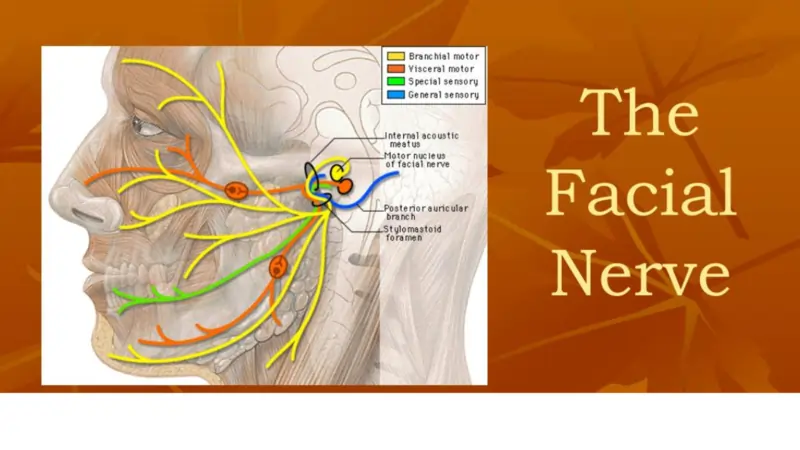
- The facial nerve arises from the Pons, it is the area of the Brainstem.
- from here, it is divided into 2 roots :
- Motor and Sensory
- The motor root is large.
- The sensory root is small.
- both the roots travel through the internal acoustic meatus.
- internal acoustic meatus is a large or long opening of the Temporal bone.
- after this, both the roots will leave the internal acoustic meatus and will enter the Facial canal.
- into the facial canal, the following will occur :
- Both sensory and motor roots will fuse together and will form one single Nerve, which is called a Facial Nerve.
- next, The Facial Nerve will form the Geniculate Ganglion, which is the collection of the nerve cell bodies.
- so, after this The Facial nerve gives rise to the following :
- Greater Petrosal Nerve :
- it innervates the parasympathetic fibers of mucous glands and lacrimal glands.
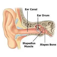
- Nerve to Stapedius muscle :
- it innervates the motor fibers of the stapedius muscle which is situated in the middle ear.
- Chorda Tympani :
- it innervates the sensory fibers to the anterior 2/3 tongue.
- and also innervates parasympathetic fibers of submandibular and sublingual glands.
- after this, the Facial Nerve will exit the Facial canal via the Stylomastoid foramen.
- The stylomastoid foramen is located posterior to the styloid process of the temporal bone.
- so this is the intracranial course of the facial nerve.
Extracranial course:
- After exiting from the cranium or skull, the Facial nerve turns superiorly and reaches up to the anterior part of the outer ear.
- From here, the Facial Nerve will divide into some motor branches. These are described here.
- Posterior Aurical Nerve :
- It provides motor innervation to some muscles around the ear.
- some motor branches will also supply to the Digastric muscle mostly, posterior muscle belly.
- and some motor branches will also supply Stylohyoid muscle.
- Now, The Main Motor Root of the facial nerve will travel continuously anteriorly and inferiorly into the Parotid gland.
- Within the Parotid gland, the nerve terminates by diving into five main branches.
- The name of these five branches are as follow :
- Temporal Branch
- Zygomatic Branch
- Buccal Branch
- Marginal Mandibular Branch
- Cervical Branch
All these branches of the facial nerve are responsible for the innervation of muscles for facial expressions.
- Branches of Facial Nerve can be simulated by putting your one side of wrist on the side of the ear, ex = Right side.
- so for simulation or identification of the right side put your right wrist on the right ear and spread your five digits like this :
- Spread your thumb over the temporal region.
- Index finger on Zygomatic bone.
- The middle finger on the upper lip.
- Ring finger on the lower lip.
- Little finger over the neck.
Functions Of The Facial Nerve:
- Functions of the Facial nerve are divided into 3 main types :
- Motor Function
- Sensory Function
- Parasympathetic Function
- all these functions are described below in detail.
Motor Function:
The first motor branch arises within the facial canal, which is :
“Nerve to Stepedius”:
function: it supplies the Stepedius muscle situated in the middle ear.
After this, there will be 3 more branches situated between the Stylomastoid foramen and the Parotid gland.
Posterior Auricular Nerve :
Function: innervates intrinsic and extrinsic muscles of the outer ear.
also supplies the occipital part of the Occipitofrontalis Muscle.
Nerve to Digasric Muscle(mostly Posterior Belly):
Function: innervates posterior belly of the digastric muscle.
Nerve to Stylohyoid Muscle :
Function: innervates Stylohyoid muscle.
After this within the parotid gland, the facial nerve bifurcates into five motor branches.
These all branches will innervate muscles of facial expression.
- Temporal Nerve :
- Function: Innervates the following muscles:
- Frontalis
- Orbicularis Oculi
- Corrugator supercilli
- Zygomatic Nerve :
- Function: innervates the Orbicularis oculi muscle.
- Buccal Nerve :
- function: innervates following muscles.
- Orbicularis Oris
- Buccinator
- Zygomaticus
- Marginal Mandibular Nerve :
- Function: innervates following muscles.
- Depressor labii inferioris
- Depressor anguli oris
- Mentalis
- Cervical Nerve :
- innervates the platysma.
Sensory Function:
The Chorda tympani branch of Facial Nerve:
Function: innervates anterior 2/3 of the tongue with the sense of taste.
Parasympathetic Function:
The Greater Petrosal Nerve and Chorda tympani branches of the facial nerve carry parasympathetic fibers of the facial nerve.
The Greater Petrosal Nerve:
- Function: Branches from pterygopalatine ganglion provides parasympathetic innervation to the following :
- Mucous glands of oral cavity, nose, and pharynx.
- Lacrimal gland.
How is the pterygopalatine ganglion formed?
- The Greater petrosal nerve arises distal to the geniculate ganglion within the facial canal.
- then the nerve will exit the temporal bone into the middle cranial fossa.
- from here, it travels across the Foramen Lacerum and combines with Deep Petrosal Nerve.
- here, they will form the Nerve to the pterygoid canal.
- The nerve to the pterygoid canal passes through the pterygoid canal.
- and will enter into the pterygopalatine fossa.
- here it will synapse with the pterygopalatine ganglion.
Chorda Tympani:
- Function: Branches from submandibular ganglion will provide parasympathetic innervation to submandibular and sublingual salivary glands.
How the submandibular ganglion is formed?
- The Chorda tympani branch combines with the lingual nerve.
- it will combine into the infratemporal fossa.
- and will form the submandibular ganglion.
Clinical Importance:
- Damage to the Facial Nerve can produce a variety of symptoms.
- it totally depends on the site of the lesion.
- there are mainly two types of lesions seen :
Intracranial lesions:
- It occurs during the intracranial course of the facial nerve, proximal to the stylomastoid foramen.
- The symptoms produced totally depend on the location or site of the lesion and on the branches that are affected.
- The most common cause of intracranial lesions is Infection.
- Mostly of External or middle ear.
- If it occurs because of an unknown cause, it is termed “Bell’s Palsy “
- In this condition, the Muscles of facial expressions will become paralyzed or will become severely weakened.
- Here is the list of branches of the facial nerve that are affected during the intracranial course.
Chorda tympani:
symptoms: Reduction in the amount of salivation.
Loss of taste of the tongue, mostly of ipsilateral 2/3.
Nerve to stapedius :
Symptoms: Hypersensitivity to sound. mostly seen ipsilateral.
The Greater Petrosal Nerve :
Symptoms: Reduction of lacrimation fluid production.
seen over the ipsilateral side.
Extracranial lesions :
- It occurs during the extracranial course of the facial nerve, Distal to the stylomastoid foramen.
- Here only the Motor functions of the facial nerve are affected.
- So it will result in paralysis or severe weakness of facial muscles.
- There are various causes of this kind of lesion, they are as follows:
Idiopathic :
Bell’s Palsy: Another name of Bell’s Palsy is idiopathic facial paralysis.it is called as an idiopathic facial palsy because the cause behind the facial paralysis may remain unknown in majority of cases.
- It might be possible reaction of a viral infection.
- It occurs because of non – suppurative inflammation of facial nerve.
- Site of facial nerve inflammation :
- Inflammation of facial nerve occurs within its canal just above the stylomastoid foramen.
- Etiology behind the disease is mostly of unknown cause.
- It can occur at any age even the gender is also not specified.
- The onset of palsy Is often sudden, the patient may have a possible history of ear ache .
- The patient wake ups with the paralysis of facial muscles , having been perfectly better the night before.
- Facial palsy is a term used to describe any kind of paralysis of facial muscles.
- Facial palsy is caused by damage to the facial nerve or cranial nerve 7.
- Which supplies the muscles of the face.
- It is categorized into two types based on the location of the casual pathology.
Central facial nerve palsy:
- Occurs due to damage above the level of the facial nucleus.
- The most common cause can be the condition of stroke.
- Here, The symptoms of facial palsy will be limited to the lower half of the face.
Peripheral Facial Nerve Palsy:
- in Peripheral facial palsy, there will be complete paralysis of one side of the face.
- so here, both the upper and lower half of the facial muscles will become affected.
Parotid gland pathology :
ex= tumor
inflammation of gland itself.
during the surgical procedure, any kind of pathology occurs.
Compression :
During forceps delivery.
because in neonates, the mastoid process is not developed fully, as it can not provide complete protection to the facial nerve.