Levator labii superioris alaeque nasi muscle
Table of Contents
Levator labii superioris alaeque nasi muscle Anatomy
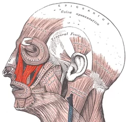
The levator labii superioris alaeque nasi muscle, often referred to as the levator labii superioris muscle, is a facial muscle located in the upper lip region. It plays a crucial role in facial expression and is part of the complex network of muscles responsible for controlling movements around the mouth and nose.
Translated from Latin, the “lifter of both the upper lip and of the wing of the nose”.
The quadratus labii, or levator labii superioris muscle, is involved in face expression and mouth and upper lip movement. Its main purpose is to elevate the upper lip, and it runs along the lateral portion of the nose. Moreover, it is involved in motions like nasal flaring, retching (vomiting), displaying oral content, and expressing disgust, grief, and contempt.
It starts on the nose’s lateral aspect and extends laterally to the zygomatic bone. The levator labii superioris is innervated by the zygomatic branch (cranial nerve VII) of the facial nerve.
Origin
Nasal bone.
Insertion
Nostril and upper lip.
Nerve Supply
Buccal branch of the facial nerve.
Together with contributions from the buccal branch of the facial nerve, the zygomatic branch of the facial nerve innervates the levator labii superioris and the other midfacial muscles between the orbicularis oculi and the orbicularis oris.
Just lateral to the levator labii superioris muscle, which is innervated from its superficial surface, is the levator anguli oris
Blood Supply
The facial artery and the infraorbital artery provide the levator labii superioris with their circulatory supplies. The angular artery, which originates from the superior labial artery and is a terminal branch of the facial artery, which is a branch of the external carotid artery, supplies the muscle directly.
The internal maxillary artery, a terminal branch of the external carotid artery, gives rise to the infraorbital artery. The infraorbital nerve, a branch of the maxillary division of the trigeminal nerve (cranial nerve V2), and the infraorbital artery pass via the infraorbital foramen.
Lymphatic Drainage
Lymphatic drainage travels through nodes in the nasolabial area, while venous outflow happens through tributaries of the facial vein that match the artery inflow.
Actions
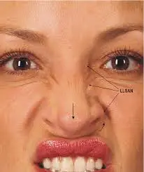
Dilates the nostril; elevates the upper lip and wing of the nose.
Structure
The levator labii superioris is a small, quadrilateral muscle that aids in eversion as well as raising the upper lip, especially when smiling. It integrates into the orbicularis oris muscle after emerging from the infraorbital border of the maxilla.
Although the muscle is more firmly attached to the skin inferior to the nasolabial fold, there is subcutaneous fat between the muscle and the dermis superior to the fold. The levator labii superioris muscle has an average length of 25 mm and a thickness of 3.5 mm.
The levator labii superioris alaeque nasi muscle, also referred to as the angular head of the levator labii superioris muscle, is located medially to the levator labii superioris. One can further separate the levator labii superioris alaeque nasi into three sections: the lip portion, the alar portion, and the furrow portion (nasolabial). There are varying amounts of superficial and deep fibers in relation to the levator labii superiors, as reported by different authors.
The frontal process of the maxilla, close to the medial palpebral ligament, is the origin of the levator labii superioris alaeque nasi. The muscle inserts into the facial soft tissue lateral to the nostril and upper lip and interdigitates with fibers of the transverse portion of the nasalis muscle.
Elevating the middle region of the upper lip, the lateral end of the nasal ala, and the upper end of the nasolabial furrow is the primary function of all three portions.
The levator anguli oris muscle, which elevates the mouth corner, is lateral to the levator labii superioris. This belly attaches on the modiolus of the orbicularis oculi after emerging from the maxilla, directly lateral to the levator labii superioris origin. This muscle is innervated from its superficial surface because its fibers extend deep to the levator labii superioris. Moreover, the zygomaticus minor muscle, which lateralizes and lifts the top lip during smiling, is connected to the lateral side of the levator labii superioris muscle. The levator labii superioris muscle is superficially located on the zygomaticus minor.
Embryology
Between the third and eighth weeks of embryonic development is when the development of facial muscles starts. The muscles begin as a thickening of the second branchial arch’s mesoderm layer. The first laminae to emerge are the occipital platysma and infraorbital lamina.
The levator labii superioris, along with numerous other face mimetic muscles, originates from both infraorbital laminae. Erroneous growth can result in congenital facial weakness, which typically manifests as segmental dysfunction matching the afflicted muscles. In contrast, traumatic or inflammatory face paralysis typically manifests as hemifacial paralysis.
Variations
According to a 2018 Taiwanese study, zygomaticus minor muscle fibers that originate in the zygomatic area and the orbicularis oculi muscle are inserted into the upper lip of 31 out of 32 adult cadavers.
Some of the zygomaticus minor fibers blended with the bottom border of the orbicularis oculi muscle in 14 (43.8%) of the cadaver specimens. The muscle is also attached to the depressor supercilii muscle, the belly of the levator labii superioris alaeque nasi, the face of the maxilla, and the palpebral ligament medially.
Clinical Importance
Treating a “gummy smile” or excessive gingival show can be done in different ways. Patients with overactive upper lip elevators that exhibit extensive gingival show respond well to botulinum toxin injections.
This is a more recent method that is far less costly and traumatic for people than doing surgery. The drawback of using botulinum toxin treatment is that it needs to be repeated because the medication’s effects wear off after three to four months, and it works best when the excessive gingival show is due to an overactive upper lip. Hyaluronic acid infiltration has also been demonstrated to be long-lasting, safe, and effective.
FAQ
Translating from Latin, the levator labii superioris alaeque nasi muscle (often reduced as alaeque nasi muscle) means “lifter of both the upper lip and of the wing of the nose”.
Superficial muscle
The superficial muscle known as the levator labii superioris (LLS) elevates and everts the top lip in concert with the other muscles of the upper lip. This muscle is the upper lip’s elevator muscle.
The top lip is raised mainly by the levator labii superioris. Stand in front of a mirror so you can see how the exercise is performed in order to concentrate on this muscle. Bring your upper lip up to your nose and purse your lips. For ten seconds, hold this posture, then let go.
References
Bloom, J. (2022, September 26). Anatomy, Head and Neck: Eye Levator Labii Superioris Muscle. StatPearls – NCBI Bookshelf. https://www.ncbi.nlm.nih.gov/books/NBK541031/

