The peroneal muscles
Table of Contents
Introduction
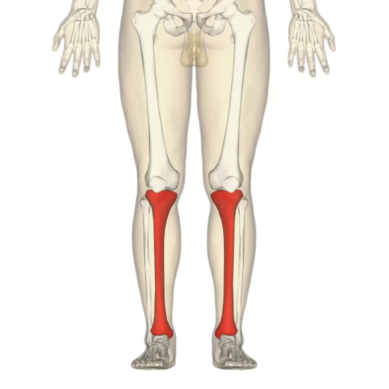
The fibular/peroneal muscles are the two muscles of the lateral compartment of the leg. These muscles are the fibularis longus and fibularis brevis.
The peroneal muscles originate from the fibula and are inserted onto the plantar surfaces of certain tarsal and metatarsal bones. These muscles play an important role in the movements of the ankle joint and support of the foot. The functions of the peroneal muscles are eversion and plantar flexion of the foot. Additionally, the peroneus longus muscle provides support to the arches of the foot.
Both peroneal muscles are innervated by the superficial fibular nerve (L5, S1), and receive their blood supply from branches of the anterior tibial and fibular arteries.
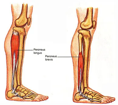
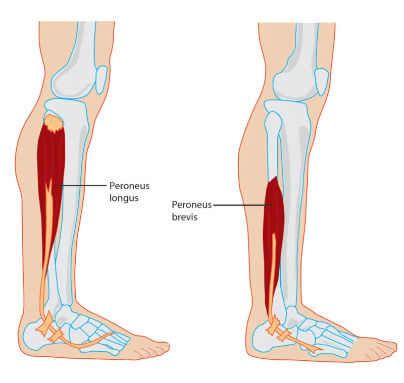
Peroneus longus
It lies superficial to the peroneus brevis muscle.
Origin:
a. Head of the fibula,
b. Upper one-third, and posterior half of the middle one-third of the lateral aspect of the shaft of the fibula
Insertion
The Peroneus longus tendon passes deep to the peroneal retinacula, runs through a tunnel in the cuboid, and is inserted into
(a) the lateral side of the base of the 1st metatarsal bone and
(b) the medial cuneiform bone adjoining part. The tendon changes its direction under the lateral malleolus and again on the cuboid bone. A sesamoid is available in the tendon in the latter situation
Nerve supply
The nerve supply of the Peroneus longus muscle is the Superficial peroneal nerve
Blood supply
The blood supply of the Peroneus longus muscle is the Anterior Tibial and Peroneal (Fibular) arteries.
Action
a. Eversion of the foot, especially when the foot is off the ground
b. It maintains the lateral longitudinal arch and transverse arch of the foot. Peroneus longus and tibialis anterior muscles are inserted into the same two bones and together form a ‘stirrup’ beneath the middle of the sole. The presence of the sling keeps the middle of the foot pulled up, and it prevents the flattening of its arches
Peroneus brevis
Lies deep in the peroneus longus muscle
Origin
a. Anterior half of the middle one-third and whole of the lower one-third of the lateral aspect of the shaft of the fibula.
b. The anterior and posterior intermuscular septa of the leg
Insertion
The Peroneus brevis tendon passes deep to the peroneal retinacula and is inserted into the lateral side of the base of the fifth metatarsal bone
Nerve supply
The nerve supply of the Peroneus brevis muscle is the Superficial peroneal nerve
Blood supply
The blood supply of the Peroneus brevis muscle is the Muscular branches of the peroneal artery.
Action
Eversion of the foot
Clinical note
When the superficial fibular nerve has been paralyzed, the eversion of the foot is severely restricted. Because of that, the supination predominates so that during the lifting of the foot, it simultaneously drifts medially (equinovarus position).
When the affected patients try to walk, it is striking how they place the lateral foot edge first in each step of the affected side. Furthermore, one may observe a shrinking of the lateral calf because of atrophy of the peroneal muscles. The most common causes of a lesion of the superficial fibular nerve include: Injuries to the fibular head, a too-tight cast or inappropriate splint positioning of the leg, and polio (infantile paralysis)
Exercise of the peroneal muscles
Strengthening the exercise of the peroneal muscles
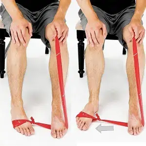
Eversion is the opposite of inversion and involves tilting the foot away from the body, bringing your little toe out and up. Eversion exercises strengthen the peroneus muscles.
Sit with legs straight in front of you. Loop a resistance band around one foot, pulling it taut against the arch.
Slowly push your foot against the resistance band, moving it toward the little toe.
Bring your foot back to a starting position.
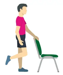
Balance on one Foot
The peroneus muscles help to stabilize the body, and balancing on one foot can help you work on the peroneal muscle.
Using the back of a chair for balance, lift one foot off the floor, keeping the other foot level and the ankle steady.
Hold for 30 seconds, then relax. Repeat 3 times on each side.
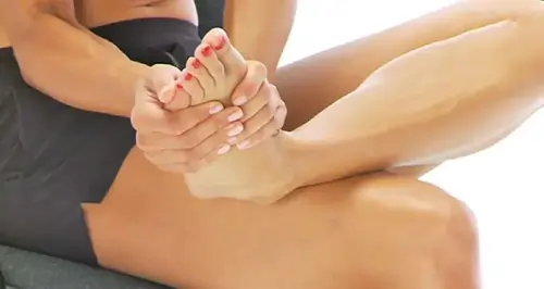
Stretching exercise of the peroneal muscles
The following exercise is a peroneus longus and peroneus brevis muscle stretch. Don’t do this exercise more often than necessary, and stop if you can feel pain.
Sit with your legs crossed with your affected foot on top.
Turn your foot inward so the sole of the foot is facing up.
Gently use your hands to apply more inward pressure for more of a stretch.
Hold for 10 to 20 seconds, then repeat three times.