Mylohyoid muscle
Table of Contents
Introduction
The mylohyoid muscle is one of the suprahyoid muscles.
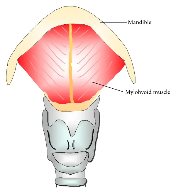
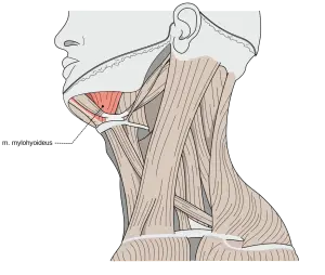
The mylohyoid muscle is a flat, triangular muscle lying deep in the anterior belly of the digastric. Both the sides of the mylohyoid muscles together form the floor of the mouth.
Along with the other suprahyoid muscles (the digastric, the geniohyoid, and the stylohyoid), The mylohyoid muscle connects the hyoid bone to the skull. The functions of this muscle are to facilitate speech and deglutition by elevating the floor of the mouth and hyoid bone and the depression of the mandible.
Origin
The mylohyoid muscle originates from the mylohyoid line of the mandible.
Insertion
The muscle fibers run medially and slightly downwards.
The posterior fibers of the muscle are inserted into the body of the hyoid bone. The middle and anterior fibers are inserted on the median raphe that unites the right and left muscles.
Nerve supply
The nerve supply of the muscle is the Mylohyoid nerve
Blood supply
The blood supply of the mylohyoid muscle is Sublingual, inferior alveolar, and submental arteries
Action
- Elevates the floor of the mouth during the first stage of deglutition.
- Helps in the depression of the mandible, and in the elevation of the hyoid bone.
Clinical significance of the Mylohyoid muscle
Conditions that can affect the mylohyoid muscle include:
- Overuse injuries
- muscle Strains
- Myopathy
- Atrophy
- Infectious myositis
- Neuromuscular diseases
- Contusions
- Lacerations
Exercise for the Mylohyoid muscle
Kiss the Sky Motion
To begin, tilt the head and look up toward the ceiling. Pucker lips mimic a kissing motion and extend the lips as far as possible. Hold the position for ten seconds, then release and lower the head. Repeat the exercise 10 times and 3 sets per day.

Tongue touches
Look up toward the ceiling and open your mouth as wide as possible. Try to touch your chin with your tongue. Hold the position for ten seconds and return to the starting position. Perform the exercise three times a day.

