Suprahyoid muscles
Table of Contents
Introduction of the suprahyoid muscles
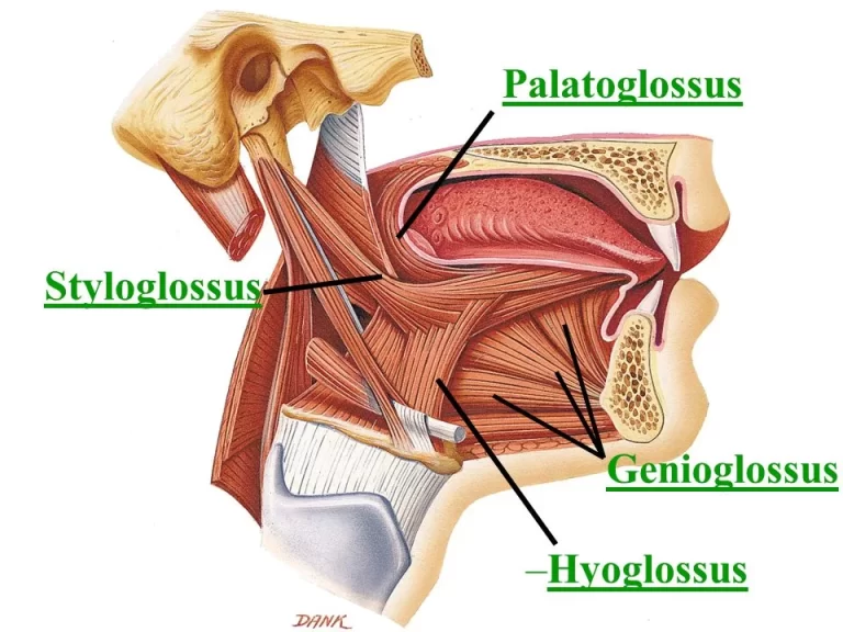
The suprahyoid muscles are the digastric, the stylohyoid, the mylohyoid, and the geniohyoid. They all are pharyngeal muscles, with the exception of the geniohyoid muscle.
They are described below.
The digastric muscle
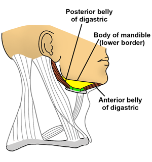
The digastric muscle lies below the body of the mandible, and extends, in a curved form, from the mastoid notch to the mandibular symphysis. It belongs to the suprahyoid group of the muscles.
This muscle is so called because it has 2 bellies. Both bellies are united by an intermediate tendon.
Origin
- anterior belly from the digastric fossa of the mandible.
- Posterior belly from the mastoid notch of the temporal bone.
Insertion
The anterior belly runs downwards and backward, and the posterior belly runs downwards and forwards to meet at the intermediate tendon. The tendon is held by a fibrous pulley which is attached to the hyoid bone. The tendon of the digastric passes between the two slips of the tendon of the stylohyoid muscle.
Nerve supply
- Anterior division by the mylohyoid nerve.
- Posterior division by the facial nerve.
Blood supply
Anterior belly of the digastric: facial artery
Posterior belly of the digastric: occipital artery
Action
- Helps to depress the mandible when the mouth is opened widely or against resistance. this action is secondary to that of the lateral pterygoid muscle.
- Elevates the hyoid bone.
The stylohyoid muscle
The stylohyoid muscle is a paired muscle located in the anterior triangle of the neck.
Stylohyoid muscle is a small slender muscle, that lies on the upper border of the posterior digastric muscle.
It is part of the suprahyoid group of the muscle which connects the hyoid bone to the mandible and skull. There is a total of four suprahyoid muscles, the other three being the mylohyoid, geniohyoid, and digastric muscles.
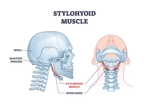
The stylohyoid muscle extends between the temporal and hyoid bones. By acting on the hyoid, it facilitates retraction of the tongue, and swallowing (deglutition), and keeps the airway open during inspiration.
Origin
The stylohyoid muscle originates from the posterior surface of the styloid process.
Insertion
The stylohyoid tendon divides into two slips that pass one on either side of the digastric tendon. The slips are attached to the hyoid bone at the junction of the body and the greater cornu.
Nerve supply
The nerve supply of the stylohyoid muscle is the facial nerve
Blood supply
The blood supply of the stylohyoid muscle is the Branches from the facial, occipital, and posterior auricular arteries
Action
- It pulls the hyoid bone upward and backward.
- With other muscles attached to the hyoid bone, it fixes the bone.
Mylohyoid muscle
The mylohyoid muscle is one of the suprahyoid muscles.
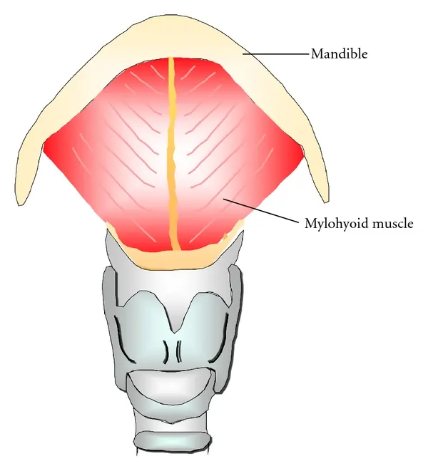
The mylohyoid muscle is a flat, triangular muscle lying deep in the anterior belly of the digastric.
Both the sides of the mylohyoid muscles together form the floor of the mouth.
Along with the other suprahyoid muscles (the digastric, the geniohyoid, and the stylohyoid), The mylohyoid muscle connects the hyoid bone to the skull. The functions of this muscle are to facilitate speech and deglutition by elevating the floor of the mouth and hyoid bone and the depression of the mandible.
Origin
The mylohyoid originates from the mylohyoid line of the mandible.
Insertion
the muscle fibers run medially and slightly downwards.
The posterior fibers of the muscle are inserted into the body of the hyoid bone. The middle and anterior fibers are inserted on the median raphe that unites the right and left muscles.
Nerve supply
The nerve supply of the mylohyoid muscle is the Mylohyoid nerve
Blood supply
The blood supply of the mylohyoid muscle is Sublingual, inferior alveolar, and submental arteries
Action
- Elevates the floor of the mouth during the first stage of deglutition.
- Helps in the depression of the mandible, and in elevation of the hyoid bone.
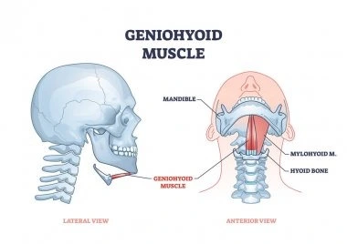
The geniohyoid muscle
This geniohyoid muscle is a short and narrow muscle that lies above the medial part of the mylohyoid muscles.
The Geniohyoid muscle is a short, paired muscle that belongs to the suprahyoid muscle group of the neck. Together with the digastric, stylohyoid, and mylohyoid muscles, it makes the floor of the oral cavity.
The geniohyoid muscle’s main function is to coordinate the movements of the floor of the mouth and the hyoid bone while swallowing and voice production.
Origin
The geniohyoid muscle originated From the inferior mental spine (genial tubercle) of the mandible.
Insertion
The geniohyoid muscle fibers run backward and downwards to be inserted into the anterior surface of the body of the hyoid bone.
Nerve supply
The first cervical nerve. the muscle fibers pass through the hypoglossal nerve.
Blood supply
The blood supply of the geniohyoid muscle is the Sublingual branch of the lingual artery
Action
- Elevates the hyoid bone.
- It May depress the mandible when the hyoid bone is fixed.
Clinical relevance of the suprahyoid muscles
Ludwig’s angina
Ludwig’s angina is a rare but serious complication from caries, tonsillitis, and gingivitis.
Ludwig’s angina occurs on the floor of the mouth and is an example of phlegmon, which is a term for inflammation of soft tissue that spreads under the skin, usually producing pus.
Bacteria spread within the connective tissue of the floor of the mouth and through the pharynx, causing symptoms such as problems in swallowing, pain in swallowing, and fever. The pathogens can migrate easily through the connective tissue of the mylohyoid muscles into the submandibular region. This may become more serious if the pathogens spread cranially into the skull or caudally into the mediastinum.
Exercise of the suprahyoid muscles
Kiss the Sky Motion
To begin, tilt the head and look up toward the ceiling. Pucker lips to mimic a kissing motion and extend the lips as far as possible. Hold the position for ten seconds, then release and lower the head. Repeat the exercise 10 times and 3 sets per day.

Tongue touches
Look up toward the ceiling and open your mouth as wide as possible. Try to touch your chin with your tongue. Hold the position for ten seconds and return to the starting position. Perform the exercise three times a day.
Hanging head lift
Lie on a bed with the head dangling over the edge. Lift the chin toward the chest. Place a hand behind the head for assistance until the jaw and neck muscles grow stronger. Hold the pose for a few seconds and slowly lower the head to its original starting position. Repeat the exercise for five to seven repetitions before relaxing.
Here is a simple exercise that can help to relieve tension in the digastric muscle. Jutting your chin forward and tilting your head slightly up, place the tips of both thumbs under your chin, one in front of the other. Next, place the tip of your tongue against the roof of your mouth, gradually increasing the pressure of your tongue while holding your thumbs firmly against the chin. Hold for ten seconds and repeat three times per day.
Another exercise is the digastric jaw protrusion resisted exercise. Push the bottom of your jaw forward against the resistance from your hand. This is a strengthening exercise for the jaw muscles, including the digastric muscles. Perform ten repetitions, three times per day.
This exercise is used to strengthen the suprahyoid muscles, including the geniohyoid. Strengthening these muscles may relieve dysphagia. This exercise has an isometric and an isokinetic portion.
Lay flat on the ground and don’t place a pillow under your head as you need it to be completely flat. Lift your head while keeping your both shoulders firmly on the ground. Tuck your chin into your chest and hold this position for 30 seconds then rest. Repeat this exercise three times.