Obturator Internus Muscle: Anatomy, Origin, Function, Exercise
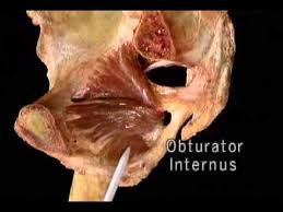
The obturator internus is a bilateral triangular-shaped muscle -the deep muscle of the gluteal region which is part of the lateral wall of the pelvis. It is seen in the superior inner side of the obturator membrane. It is also called The internal obturator muscle.
This muscle is mainly a part of the lower limb muscles. Together with other external rotators like the piriformis, quadratus femoris, superior gemellus, and inferior gemellus muscles, it includes the deep layer of muscles of the gluteal region, located under the inferior half of the gluteus maximus muscle.
The obturator internus with Superior and inferior gemellus muscle form the triceps coxae, which act together on the hip, mainly through external rotation and extension of the thigh.
Table of Contents
Obturator internus muscle anatomy:
Obturator internus muscle origin, insertion, and nerve supply, blood supply, action are discussed in this article.
Origin:
- the pelvic surface of the obturator membrane.
- the pelvic surface of the body of the ischium, ischial tuberosity, ischiopubic rami, and ilium below the pelvic brim.
- obturator fascia.
Insertion:
the tendon of the obturator internus leaves the pelvis through the lesser sciatic foramen. here it bends at a right angle around the lesser sciatic notch and runs laterally to be inserted into the medial trochanter of the femur.
Nerve supply:
nerve to obturator internus (l5-s1).
Blood Supply:
The main blood supply of the Internal obturator muscle is the Obturator artery; internal pudendal artery
Action:
Abducts & laterally rotates the extended hip, abducts the flexed thigh at the hip, and stabilizes the hip during walking.
Relations
The obturator internus muscle is located in the pelvis area. Here, the medial side of the obturator muscle is located obturator fascia, whose central thickening provides the attachment to the muscles of the pelvic diaphragm muscles (levator ani).
The fascia of the muscle is medially located to the obturator artery and nerve, as they travel anteroinferiorly from the anterior trunk on the lateral pelvic wall to the upper part of the obturator foramen. The gluteus maximus muscle and the ischial nerve are located superficial (posterior) to the muscle.
Obturator internus exercise:
There is mainly two types of exercise:
- Stretching exercise
- strengthening exercise
Obturator internus muscle stretch
If your obturator internus muscles are tight, here are two easy-to-perform home exercises for a release of muscle. The following exercise is the best exercise for obturator internus muscle stretch.
Frog pose exercise:

This is the best obturator internus muscle stretch exercise.
- On a soft mat you can start with prone all position (quadruped) while keeping your both knees wide apart and your feet to the sides. you should place your both hands flat on the floor, nearly shoulder-width apart.
- Your hands keep under your shoulders, and your knees under your hips. Make your knees bent and your ankles behind you, in line with your knees, Turn your toes out to the sides.
- Then gradually start to push your knees out to the sides.
- When you start to feel the stretch on your inner thighs and groin, stop pushing and take a deep breath again. Hold for 10 to 20 seconds and then return to the first position. Repeat this exercise 5 to 10 times.
Supine frog stretch:
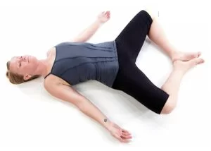
- you start with a supine position on a soft mat with your legs slightly flexed and keep your arms at your sides.
- The soles of your feet keep together and pull your heels as close to your groin as possible gradually as you feel stretch at your hip area.
- Hold this stretching for 10 to 20 seconds.
- Then bring your knees together and up which will push your feet to the floor and hold that position for 10 to 20 seconds.
- return to normal position and extend your legs to repeat the exercise.
- Perform 10 to 12 repetitions.
How to strengthen the obturator internus?
The most common exercises that help to strengthen your lower limb with the use of body weight are running, walking, stair climbing, squats, lunges, and jumping exercises are also strengthen the obturator internus muscle. This exercise helps you maintain your body balance and hip joint stability to prevent falls, injuries, and movement.
Full squatting:
This exercise strengthens your limb muscles with also strengthens the obturator internus. This is best to perform a home exercise where no equipment is required.
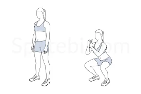
You can start with a standing position, keep your feet width apart and firm on the floor, and your toes are laterally rotated, your knees are straight, and your back is also straight, you can now start squatting gradually down. Your ankles, knees, and hips will bend in simultaneously while keeping your back straight.
As you start to lower, your knees will go forward over your toes, and your hips will go backward to keep your body weight centrally.
At the same time, Your trunk and hip will stay neutral position and straight as you bend at your hips. Try to keep your pelvis will remain in a neutral position, without tilting forward or backward.
The full squat exercise helps to strengthen your hips, knees, ankles-related muscles, and even your back muscles and also improves the strength of other related muscles mainly quadriceps, obturator internus, Glutei, and calf muscles, and also related spinal muscles.
Variations
Only a few physiological changes in the obturator muscles have been identified in the literature. To better understand piriformis preservation following posterior hip arthroplasty, Pine et al. looked at differences in the connection between the piriformis and obturator internus in one research. An analysis was conducted on the tendon crossing angles, the insertion position onto the greater trochanter, and the fusion between tendons before insertion. It was discovered that the piriformis and obturator internus tendons joined before implantation on the femur in 10% of patients.
It has also been observed that ageing has a specific impact on the architectural design of the obturator internus, which is somewhat connected to anatomical variances. Research indicates that aging-related modifications to the obturator internus’s structure resulted in a decline in the muscle’s capacity to generate force and an increase in fibrosis.
Clinical importance:
Obturator internus muscle strains
Obturator internus muscle strains are rarely seen in injury, reported mainly in high-intensity sports. Overuse of repetitive sports injuries is the main cause of sports injuries. Symptoms mainly are buttock pain and tenderness. Pain increase when you start playing sports like kickball or other activity where muscle action is required. Treatment mainly depends upon the severity, mostly use of the RICE Principle – Rest for 2 to 3 weeks and use of Ice packs, Elevation of the Limb if swelling is seen and use of a crepe Bandage or Rigid Taping / Kinesio may be helpful.
After 3 weeks regular pain-free strengthening and stretching exercises help you return to sports.
The obturator internus has demonstrated its function as a hip stabilizer influencing the effect of the posterior approach in total hip replacement (THR) in the study conducted by Solomon et al. (2010). The most popular technique for total hip replacement is the posterior approach, which involves cutting off the piriformis, obturator externus, and obturator internus tendons (short external rotators) as they penetrate into the greater trochanter. The incidence of hip dislocation following surgery is greater even when the tendons have been repaired because the short external rotators also function as hip stabilizers. Therefore, while doing THR, different procedures are preferred over the posterior approach.
Gemelli-obturator syndrome
When the sciatic nerve is pinched during passive hip rotation between the obturator internus and the gemellus muscles, it might result in gemelli-obturator syndrome.
Physical therapy, gabapentin, non-steroidal anti-inflammatory medications, muscle relaxants, and rest are among possible treatments for this illness.
Posterior Hip Dislocation
The obturator muscles are clinically significant when there is a hip injury, such as a dislocation, because of their close proximity. The most prevalent kind of hip dislocations are posterior ones. They usually occur when the hip is flexed and adducted and a posteriorly directed force is given to a flexed knee. This dislocates the hip by driving the femoral head posteriorly. Gluteal muscles and short external rotator muscles, including the obturator internus, may sustain damage during dislocation. But, the most frequent side effects of posterior hip dislocation are damage to the sciatic nerve and avascular necrosis. The femoral head’s main blood supply is the deep branch of the MCFA.
The risk of avascular necrosis is significantly increased when damage is done to this artery. Research has indicated that the obturator externus is essential for safeguarding the deep branch of the MCFA in the event of a hip dislocation in either direction. Hip stabilizer muscles are tracked in gait analysis and muscle rehabilitation following posterior hip dislocation.
FAQs
Although the obturator nerve is located deep within the pelvis, pregnancy, accidents, and surgical trauma can all affect it. Muscle weakness and discomfort in the medial thighs are signs of obturator neuralgia.
The anterior divisions of the sacral plexus give birth to the nerve that supplies the obturator internus. It originates from the L5-S2 nerve roots and leaves the pelvis through the larger sciatic foramen, which is located inferior to the piriformis muscle and usually lies between the pudendal nerve & the posterior cutaneous nerve
References
Larson, M. R. (2023, January 17). Anatomy, Abdomen and Pelvis, Obturator Muscles. StatPearls – NCBI Bookshelf. https://www.ncbi.nlm.nih.gov/books/NBK589636/





4 Comments