Masseter Muscle Anatomy
Table of Contents
Introduction
The masseter is a quadrilateral muscle that covers the lateral surface of the ramus of the mandible. The masseter muscle fibres are arranged in three layers.
The masseter muscle is a paired, strong, thick, and rectangular muscle that originates from the zygomatic arch and extends down to the mandibular angle. It contains a superficial and a deep part.
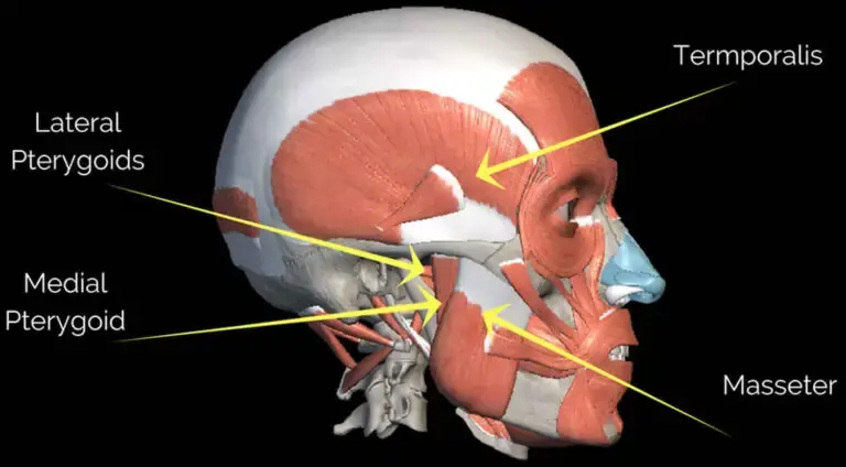
It is one of the mastication muscles, a group of muscles that also includes the temporal muscle, lateral pterygoid muscle, and medial pterygoid muscle. The masseter muscles specific functions are elevation and protrusion of the mandible, as well as providing support to the articular capsule of the temporomandibular joint.
Origin of Masseter muscle
Superficial layer from the anterior 2/3 of the lower border of the zygomatic arch and from the process of zygomatic of the maxilla
Middle layer from the anterior 2/3 of the deep surface and posterior 1/3 of the lower border of the zygomatic arch.
Deep layer from the deep/inferior surface of the zygomatic arch (posterior 1/3)
Insertion of masseter muscle
The superficial fibres pass downwards and backwards at an angle of 45 degrees. superficial fibres are inserted into the lower part of the lateral surface of the ramus of the mandible.
The middle and deep fibres pass vertically downwards. the middle fibres are inserted into the middle part of the ramus, and the deep fibres into the upper part of the ramus and into the coronoid process.
Nerve supply
The nerve supply of the masseter muscles is the Masseteric nerve, a branch of the anterior division of the mandibular nerve (CN V3)
Blood supply
The blood supply of the masseter muscle is the Masseteric artery
Action of Masseter muscle
The masseter muscle Elevates the mandible and clenches the teeth
Clinical significance
Infections and submasseteric abscesses
Infected with the bacteria Clostridium tetani, strong and persistent spasms of the masseter muscle may occur. That kind of contraction is called trismus and may interfere with the normal feeding process.
Since the masseter is involved with submasseteric space, it has an important role when it comes to submasseteric abscesses. These abscesses are rare, so they are easily confused with parotid gland infections. The origin of the infection is commonly odontogenic, from pericoronitis in the mandibular third molar, especially when the apices of the tooth lie very close to or within the space. As a result, the infection typically spreads easily to this area where these abscesses tend to be chronic.
A major hallmark of a submasseteric space infection is the upper mentioned trismus, so the masseter muscle is actually sending signals that the infection is all about submasseteric space instead of any surrounding structures.
By knowing the relations and functions of the masseter muscle, we can easily narrow down our differential diagnosis options for the patient who is suffering from some form of facial lesions.
Exercise of the masseter muscle
The exercise starts by resting the chin on the tip of the thumb. Open the mouth while applying light consistent pressure with the thumb. Continue holding the mouth open while applying pressure for 10 to 20 seconds and then close the mouth. Repeat this exercise ten to fifteen times and three to five sets per day.
Side-to-side Exercise
Place a pen or pencil in the mouth and hold it between your teeth. Slowly move the jaw from one side to the other side. Repeat this exercise ten to fifteen times and three to five sets per day.
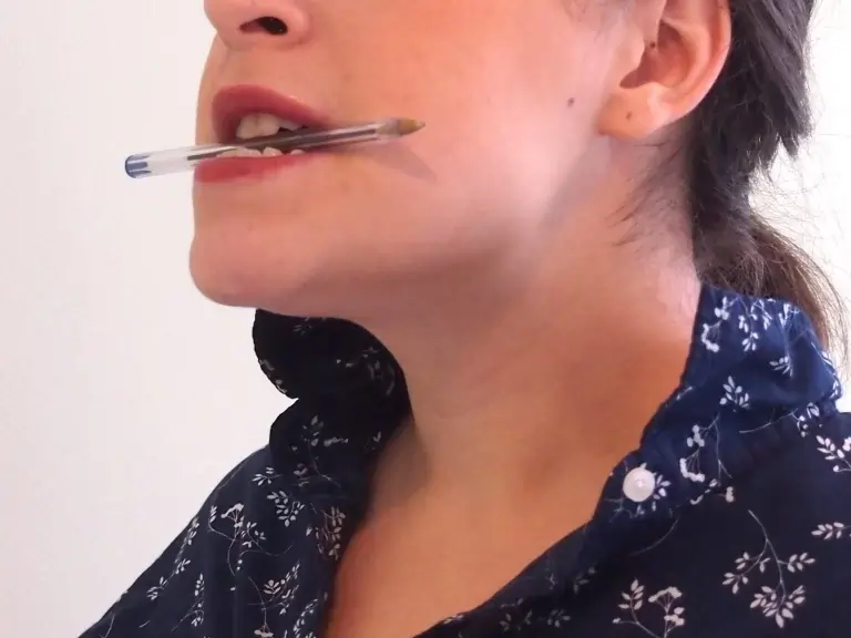
Resisted opening of the mouth
Place your thumb under your chin. Open your mouth slowly, pushing gently against your thumb for resistance. Hold for three to five seconds, and then close your mouth.

Resisted closing of the mouth
For this exercise squeeze your chin with your index and thumb with one hand. Close your mouth as you place gently resistance on your chin. This will help to strengthen your muscles that help you chew.


3 Comments