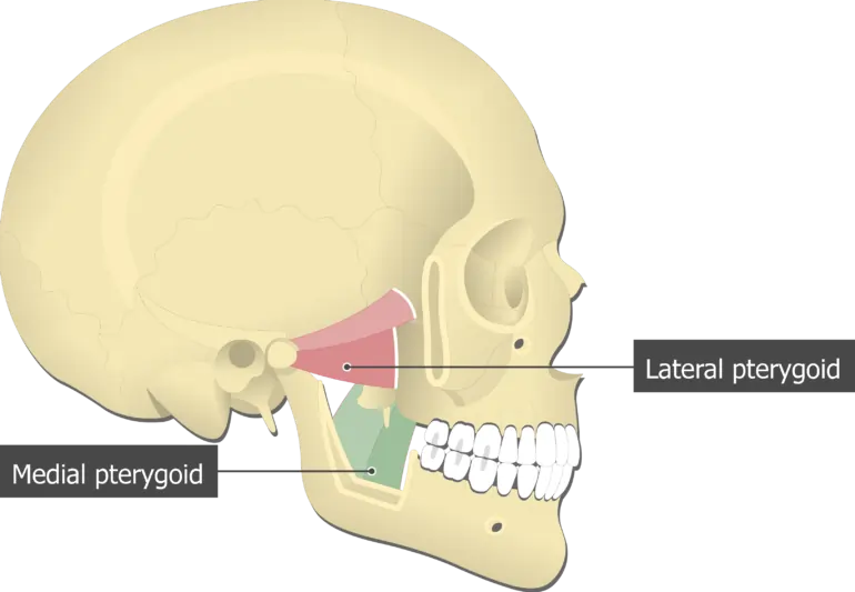
Medial pterygoid muscle Anatomy
Table of Contents
Introduction
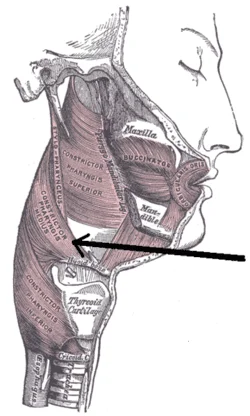
The medial pterygoid is a thick, quadrilateral muscle of the face and it has a small superficial head and a large deep head which forms the major part of the muscle.
The medial pterygoid muscle is supplied by the mandibular branch of the trigeminal nerve (V). It is important in mastication (chewing).
It belongs to the group of mastication muscles, along with the lateral pterygoid muscle, masseter, and temporal muscles.
Although having different origins of both heads, both heads insert into the inner surface of the mandible, creating an axis for a strong pull of this bone. Unilateral contraction of medial pterygoid muscle causes rotation of the mandible, while bilateral contraction causes elevation and protrusion.
Origin of Medial pterygoid muscle
The superficial head(small slip) of the medial pterygoid muscle originates from the tuberosity of the maxilla and adjoining bone.
The deep head of the medial pterygoid muscle originates from the medial surface of the lateral pterygoid plate and adjoining part of the palatine bone.
Insertion of Medial pterygoid muscle
The medial pterygoid muscle fibers run downwards, backward, and laterally to be inserted into the roughened area on the medial surface of the angle and the adjoining part of the ramus of the mandible, below and behind the mandibular foramen and the mylohyoid groove.
Nerve supply
The nerve supply of the medial pterygoid muscle is the Branch of the main trunk of the mandibular nerve.
Blood supply
The blood supply of the medial pterygoid muscles is the pterygoid and buccal branches of the maxillary artery. To a lesser extent, ascending palatine artery and muscular branches of the facial artery also contribute to the blood supply of the medial pterygoid muscle.
The action of Medial pterygoid muscle
Elevates the mandible
Helps to protrude the mandible
See “c” under lateral pterygoid
Clinical significance
The medial pterygoid muscle can sometimes be injured during inferior alveolar nerve block due to it being in close proximity to the nerve. The injury occurs if the anesthetic needle is placed too medially and accidentally injected into the medial pterygoid muscle instead of the inferior alveolar nerve.
This may cause hemorrhage and the development of medial pterygoid trismus hours to days after the procedure. This manifests in the inability to open the mouth completely and significant medial pterygoid muscle pain when attempting to open the mouth beyond the restriction.
Exercise for the medial pterygoid muscle
Open wider (Mouth opening exercise)
Start with slowly opening your mouth as wide as possible. If clicking or popping of the jaw occurs, immediately discontinue stretch. Hold the stretch for ten to fifteen seconds then close your jaw. Repeat this stretch five to seven times.

Tongue Stretches
Move the tongue to where it rests on the back of your upper front teeth. then move the tongue back until it is resting on the soft tissue of the roof of the mouth. Open the mouth and make sure to keep the tongue in the same position. Hold for approx ten seconds and return to the starting position. Complete three repetitions of these exercises.
To actively stretch the medial pterygoid, the patient is in the supine position and places two fingers behind the lower incisor teeth and the thumb under your chin, and by pulling the mandible forward and downward, the patient opens the jaw fully. The opposite hand is placed on the forehead to stabilize your head and neck.