Temporalis Muscle Anatomy
Table of Contents
Introduction of the Temporalis muscle
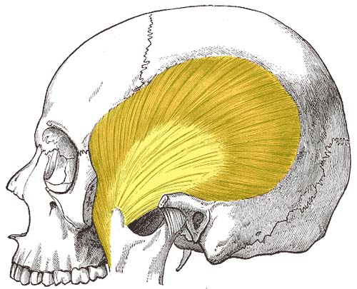
In anatomy, the temporalis muscle is also known as the temporal muscle. This muscle fills the temporal fossa.
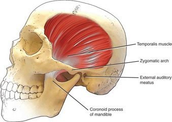
The temporalis muscle is a thin, fan-shaped muscle located within the temporal fossa of the skull. Along with the medial pterygoid muscle, lateral pterygoid, and masseter muscles, it belongs to the group of mastication muscles. The temporalis muscle runs superficially, from the temporal bone to the coronoid process of the mandible.
The main function of this muscle is to produce the movements of the mandible at the temporomandibular joint(TMJ) and thus facilitate the act of mastication. Its anterior portion moves the mandible in elevation while its posterior fibers pull the mandible in the retrusion.
Origin of Temporalis muscle
Temporal fossa, excluding the zygomatic bone,
Temporal fascia
Insertion of Temporalis muscle
The fibers of the temporalis muscle converge and pass through the gap deep to the zygomatic arch. They are inserted into The margins and deep surface of the coronoid process; and the anterior border of the ramus of the mandible.
Nerve supply of Temporalis muscle
The nerve supply of the temporalis muscles is the Deep temporal branches from the anterior division of the mandibular nerve (CN V3)
Blood supply of Temporalis muscle
The blood supply of the temporalis muscle is the Deep temporal branches of the maxillary artery, middle temporal branches from the superficial temporal artery
The action of Temporalis muscle
Anterior fibers of the temporalis muscle: Elevation of the mandible
Posterior fibers of the temporalis muscle: Retraction of the mandible
Clinical significance of the Temporalis muscle
The tension of the temporal muscle can cause pain in the temporal area. Common causes include:
misalignments of the teeth and jaws
trauma
a prolonged immobilisation (e.g. after a mandibular fracture)
teeth grinding (bruxism)
a dental intervention during which the patient’s mouth had to be open for a long period of time.
inflammation of the superficial temporal artery, which runs in front of the ear along the zygomatic arch to the temporal region. Vasculitides, such as giant cell arteritis, frequently involve the superficial temporal artery and cause swelling and pain in the temporal area. The diagnosis can be confirmed by a biopsy of the temporal artery.
Seizure
A myotendinous rupture of the temporalis muscle may occur during a seizure due to extreme clenching of the jaw. During a seizure, the contralateral temporalis muscle can enter spastic paralysis, this clenching in extreme cases can lead to a rupture specifically on the myotendinous insertion on the coronoid process of the mandible.
Surgery
The temporalis muscle can be used in reconstructive surgery of the mouth.
While pterional craniotomy, incisions are often chosen based on the ease of reattaching the temporalis muscle after the bone fragment is replaced.
Exercise for the Temporalis muscle
Clenched your teeth tightly and hold for 10 seconds. Next, clench down on your back teeth for a count of 10 seconds, feeling your temporalis flexing with each clench. Relax. Repeat this exercise three times.

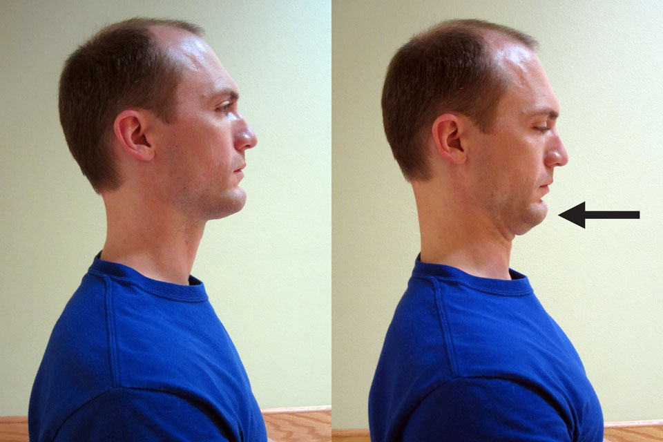
Jaw Tamporo Mandibular Joint retraction
Using your fingers, push the bottom of your jaw backward towards your throat.
Place the both pad of the thumb of your hands just above the cheekbones and press in.
While slowly opening the mouth, press in against the temporalis with the thumbs. Slowly slide the thumbs up and then along the muscle.
End the compression and stretching when you meet the edge of the muscle, which is about 1 to 2” above the ear.
Repeat at least 10 times.
Be sure to address tension in all parts of the muscle.
You can apply some lotion to the face may make the stretch easier and more comfortable.