
Pronator Quadratus Muscle
Table of Contents
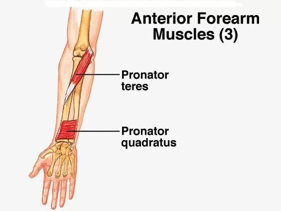
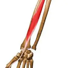
Pronator Quadratus Muscle Anatomy
The Pronator Quadratus muscle is a small, square-shaped muscle located in the forearm of the human arm. It plays a crucial role in forearm pronation, which is the rotational movement of the forearm that turns the palm of the hand downward or backward.
- Pronator quadratus is a quadrangular, thin, short, and flat muscle lying within the anterior compartment of forearm.
- It is part of the deep group of forearm flexors, together with flexor digitorum profundus and flexor pollicis longus.
- These three muscles are overlaid by the superficial group of forearm flexors.
Origin
- Medial, anterior surface of the ulna.
Insertion
- Anterior surface of the radius.
Nerve supply
- Median nerve
The anterior interosseous nerve of the forearm innervates the pronator quadratus, mostly with input from the C7 and C8 spinal neurons. The median nerve, which emerges from the brachial plexus, branches off to become the anterior interosseous nerve.
Actions
- Pronates the forearm
The lateral side of the radius is pulled toward the ulna when the pronator quadratus contracts, pronating the hand. The two bones in the forearm are held together by its deep fibers. Additionally, this muscle may not be present in some persons, but this has little impact on pronation because the pronator teres plays a big part in it.
Blood Supply
- Anterior interosseous artery.
The anterior interosseous artery, which branches off of the common interosseous artery, supplies arterial blood to the pronator quadratus.
Length
- The mean length of the muscle is 6 cm.
Width:
- The mean width of the muscle is 3.5 cm.
Structure
From the most distal quarter of the anterior ulna to the distal quarter of the radius, its fibers run perpendicular to the direction of the arm. It has two heads: the superficial head inserts into the anterior distal diaphysis and anterior metaphysis of the radius, coming from the anterior distal portion of the diaphysis (shaft) of the ulna.
Similar in origin, the deep head enters close to the ulnar notch. It is the only muscle that has a single ulnar and radial attachment at each end. The anterior interosseous artery is the source of arterial blood.
Muscle Relations
The pronator quadratus is the muscle which is located deepest within the anterior (flexor) compartment of the forearm. As a result, it is situated beneath the flexor digitorum profundus and flexor pollicis longus, the other deep forearm flexors. The pronator quadratus, which is situated distally in the forearm, superficially covers the interosseous membrane.
As it travels from the anterior to the posterior compartment of the forearm, the anterior interosseous artery punctures the interosseous membrane proximal to the pronator quadratus. Along its path to the palmar arch, a branch of the anterior interosseous artery also descends deeply to the pronator quadratus. The palmar carpal branch of the radial artery arises near the muscle’s distal border, and it goes anteriorly to the pronator quadratus. The pronator quadratus’s deep surface is reached via the anterior interosseous nerve’s posterior path.
Clinical Significance
Myofascial Trigger point (TrP)
There are two primary referred pain patterns in the Pronator Quadratus muscle. The medial aspect of the forearm most frequently feels painful both proximally and distally. Many times, the discomfort will radiate from the medial epicondyle to the fifth finger of the hand. The third and fourth fingers were also affected, which is the second most typical pattern of pain.
Pronator Spasticity
In order to treat pronator spasticity in stroke patients, the motor point of the pronator quadratus is injected with neurolytic drugs like Botulinum toxin, phenol, or alcohol.
Anterior Interosseous Nerve (AIN) lesions
The Pronator Quadratus muscle, which serves as the main muscle for this diagnostic method, is used to make an electrophysiologic diagnosis of AIN lesions.
Distal Radius Fracture
The most common approach for treating this fracture is volar plating. A potential side effect of this method is flexor tendon rupture, which can be avoided by repairing the pronator quadratus muscle during surgery.
In the early postoperative period, better pronation movement, better radioulnar joint stability, good grip, and reduced pain can all be attributed to fixing the distal radius fracture while maintaining the pronator quadratus muscle.
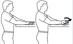
Stretching Exercise for pronator quadratus muscle:-
- Sit with forearm supported and elbow bent.
- Palm should be up.
- Grasp thumb with other hand and pull down while twisting wrist to thumb side, as shown.
- Keep thumb near base of index finger.
- Hold, relax and repeat.
- Hold end position for 10-15 seconds
- Repeat for 5-6 times
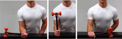
Strengthening Exercise:
- Muscle can be strengthen using theraband, flex-bars, weight cuffs, dumbells.
- First, place the hand in supination and grab the end pf flex-bar and then pronate the forearm.
- Resistance is used depending upon the condition of patients/clients.

FAQs
The lateral side of the radius is pulled toward the ulna when the pronator quadratus contracts, pronating the hand. The two bones in the forearm are held together by its deep fibers.
The pronation of the radioulnar joint is caused by the pronator teres and pronator quadratus muscles. The radioulnar joint pronates as a result of this muscle’s contraction, which drags the distal end of the radius over the ulna.
5 Comments