
Soleus Muscle
Table of Contents
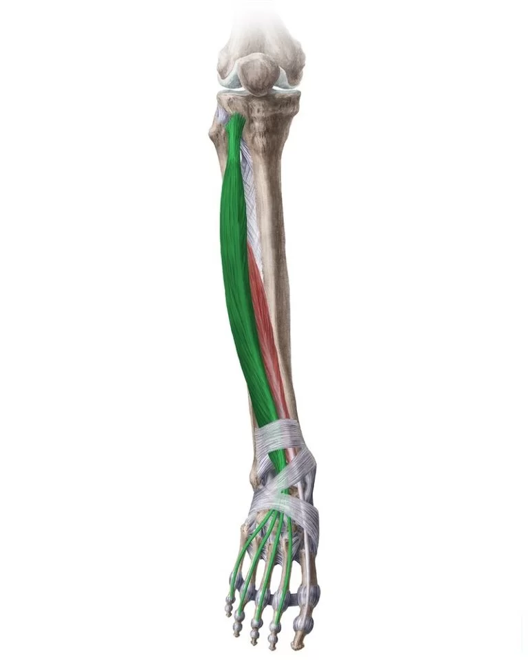
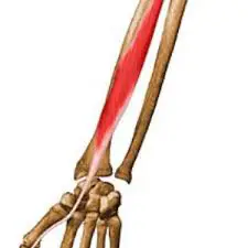
Soleus muscle Anatomy
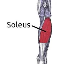
The soleus muscle is a wide flat leg muscle found on the posterior leg. It runs from just below the knee to the heel and lays immediately deep into the gastrocnemius.
These two muscles, along with the plantaris muscle, belong to the group of superficial posterior compartment calf muscles.
They make up the trio of muscles known as the triceps surae, together with the gastrocnemius. They both function in numerous fundamental movements, including walking, running, and leaping, and they both insert on the calcaneus via the calcaneal tendon. The triceps surae muscle bellies’ size and shape affect the interindividual variations in human calves’ appearance, which ranges from slender to rather significant.
Origin
The soleus muscle arises from the soleal line on the dorsal surface of the tibia, medial border of the tibia, head of the fibula, and posterior border of the fibula.
Insertion
Soleus insert onto the posterior surface of the calcaneus via the calcaneal tendon. The calcaneal tendon, commonly called the Achilles tendon
Nerve supply
The soleus is innervated by the ventral rami of S1 and S2 spinal nerves, carried by the tibial nerve into the posterior compartment of the leg.
Blood supply
The superior branch arises from the popliteal artery while the inferior branch arises from a peroneal artery (fibular artery) or the posterior tibial artery.
Actions
Soleus contraction causes strong plantar flexion.
These strong muscles are essential for running, walking, and maintaining equilibrium. The soleus muscle is crucial for preserving upright posture; without it, the body would tip over front.
Additionally, while in an upright position, the soleus, also known as the skeletal muscle pump, peripheral heart, or sural (tricipital) pump, is in charge of returning venous blood from the periphery to the heart.
Compared to many other muscles, the soleus has slower muscle fibers. Some animals, including cats and guinea pigs, have soleus that are made entirely of sluggish muscle fibers. The proportion of slow fibers in human soleus varies, ranging from 60% to 100%.
The soleus, often known as the first gear muscle, is the muscle most useful for plantarflexion when the knee is bent. Because the gastrocnemius muscle originates in the femur, its effective tension is limited when the leg is bent. Because its slow-twitch fibers withstand exhaustion, the soleus is the major muscle used for plantarflexion during regular activity (e.g., walking).
Relations
The soleus muscle, which is located beneath the gastrocnemius, is closer to the skin, being superficial to it.
Between the two muscles lies the plantaris muscle, along with a section of its tendon.
The transverse intermuscular septum, which divides the leg’s superficial and deep posterior compartments, is located deeper into it (away from the skin).
The tibialis posterior muscle, flexor digitorum longus, flexor hallucis longus, posterior tibial vein, posterior tibial artery, and tibial nerve are located on the other side of the fascia.
The posterior compartment is the bulge of muscle medial to the tibia on the anterior side of the leg whereas the anterior compartment is lateral to the tibia. In the superficial center of the tibia is the soleus.
Synergists
Gastrocnemius
Plantaris
Tibialis posterior
Peroneus longus and Brevis
Flexor Digitorum Longus (FDL)
Flexor Hallucis Longus (FHL)
Antagonists
Tibialis anterior
Palpation
To make sure the gastrocnemius stays relaxed when palpating the soleus, plantarflex the ankle while flexing the knee to 90 degrees. The Achilles tendon’s lateral and medial sides can then be used to palpate the lateral and medial portions of the muscle. The majority of the muscle’s distal to proximal distance can be felt with palpation; however, if the gastrocnemius heads are large, it will be more challenging to feel the proximal attachments.
Accessory soleus muscle (ASM)
Between 0.7 and 5.5% of people have it. It typically manifests in the second or third decade of life and is 2:1 more common in girls than in males. For the most part, it is unilateral.
This supernumerary muscle is situated in the posterior upper third of the fibula, between the posterior portion of the tibia and the fibular head, in the oblique soleus line, underneath the gastrocnemius muscle. The ASM extends anteriorly and medially from its point of origin to the Achilles tendon.
Soleus muscle Strengthening exercise:
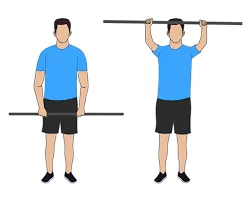
Double-Leg Calf Raise:
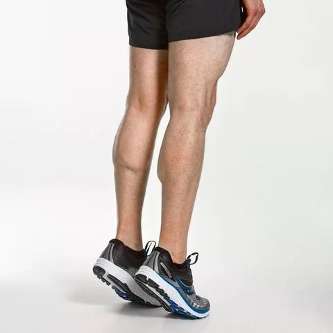
Calf raises are the classic calf-strengthening exercise. They use your body weight to strengthen and tone the gastrocnemius and soleus.
Starting position: Stand near a wall for balance. Place your feet hip-width apart, and make sure your ankles, knees, and hips are in vertical alignment to protect your joints.
Action: Press down into the balls of both feet to raise your body upward. Keep your abdominal muscles pulled in so that you move straight upward, rather than shifting your body forward or backward.
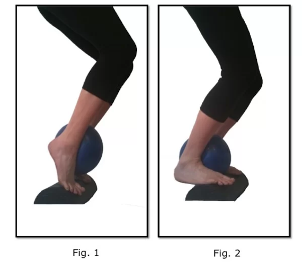
Single-Leg Calf Raise:
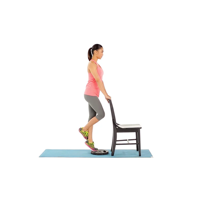
You can increase the intensity of the calf raise by doing it on one leg. That way you can strengthen your calf muscles even more.
Starting position: Stand on one leg near a wall for balance with the other leg bent behind you. Be sure the ankle, knee, and hip of the leg you’re working are in vertical alignment to protect the joints.
Action: Press down into the ball of your foot to raise your body upward. Keep your abdominal muscles pulled in so you avoid shifting forward or backward.
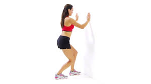
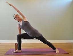
Stretching exercise of Soleus muscle:
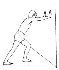
- Stand about arm’s-length from the wall.
- Lean forward and place both hands on the wall about shoulder-width apart.
- Extend one foot (the side to be stretched) behind you with one heel on the ground, and the other foot closer to the wall.
- Lean into the wall with your hips until you feel a stretch in the calf of the extended leg.
- Hold this stretch for about 30 seconds, and then change sides.
- For a deeper stretch, move your foot farther back.
- This stretch is similar to the Achilles tendon and heel stretch; however, by keeping your knee straight, you focus the stretch on the calf rather than the Achilles tendon.
Clinical Importance:
Deformity:
Clubfoot is a birth defect where one or both feet are rotated inward and downward. The affected foot and leg may be smaller in size compared to the other. Approximately 50% of cases of clubfoot affect both feet. Most of the time, it is not associated with other problems. Without treatment, the foot remains deformed, and people walk on the sides of their feet. This may lead to pain and difficulty walking.
ACL Injury and Soleus Muscle:
A better understanding of the agonistic behavior of the soleus muscle on the anterior cruciate ligament may lead to the development of training and rehabilitation strategies that could reduce the incidence of injury and improve function in both patients with anterior cruciate ligament deficiency and patients who have undergone anterior cruciate ligament reconstruction.
Leg muscles have extensive fascia surrounding them, which makes them vulnerable to compartment syndrome. This condition is related to inflammation of the tissue that compresses nerves and alters blood flow. If compartment syndrome is not treated, it can result in neuropathy, blood clots, and muscular atrophy.
Soleus tear
In sports medicine and primary care clinics, strained calf muscles are often observed injuries. Overuse is a common cause of a soleus tear. Runners often sustain injuries as a result of overtraining and exhaustion. Because long-distance and endurance running and walking demand continuous soleus motion, athletes who specialize in these sports are more vulnerable to injury.
Some of the symptoms are:
Ache in the lateral leg on either side of the soleus
Calf weakness and poor plantar flexion, particularly when the knee is bent; dull calf pain from exercise; pain from foot dorsiflexion or pressure on the calcaneal tendon.
The RICE approach (rest, icing, compression, and elevation) is part of the treatment. It’s important to distinguish between a gastrocnemius tear and a soleus tear, as the latter is typically the result of abrupt trauma from rapid motions like jumping or running. The Cause of it has led to it being referred to as the tennis leg.
Soleus pain
A Soleus tear or overuse may be the cause of soreness in the soleus. Affected individuals usually claim that their pain starts during exercise and becomes better over time. Usually, the pain returns after the training. The lateral middle area, the head of the muscle, which may transmit pain all the way up into the sacroiliac area, or the calcaneal tendon and muscle belly transition are the three primary places where muscle discomfort usually arises. It may not always be easy to diagnose a soleus injury. To identify diseases inside the muscle, MRI or ultrasound are typically utilized in addition to clinical symptoms and physical examination.
A deep vein thrombosis (DVT), which poses a danger of pulmonary embolism (PE) and is a potentially fatal medical emergency, may also be the cause of sudden calf pain. In addition to calf pain, other common signs of DVT include calf swelling and redness. Heparin, warfarin, and factor Xa inhibitors are examples of pharmacotherapeutic drugs used in anticoagulation therapy. One preventive tactic is to use compression stockings.
FAQ
Because of its similar function to the heart’s within the circulatory system, the calf muscle (soleus) is frequently referred to as the “second heart.” Blood can travel throughout the body with just one heartbeat, arriving to the lower extremities in a matter of seconds.
The soleus is a deep muscle located in the calf that is responsible for plantar flexion, which is pointing the toes downward. It is an important muscle for walking, running, and jumping, and it can also help to improve balance and stability.
Here are some exercises that you can do to strengthen your soleus:
Standing calf raises: Stand with your feet shoulder-width apart and your toes pointing forward. Slowly raise your heels off the ground, then slowly lower them back down. You can make this exercise more challenging by holding onto a wall or railing for balance. Repeat 10-15 times for 3 sets.
The soleus muscle is useful for walking, dancing, and running. It also aids in keeping the body from falling forward. The soleus muscle aids in maintaining balance while you stand on one foot. It is a strong muscle that continues to contract during all kinds of weightlifting exercises.
When compared to gastrocnemius injuries, soleus strains also typically manifest clinically as subacute injuries rather than as severe ones. Calf tightness, stiffness, and pain that gets worse over several days to weeks is the classic appearance. Jogging or walking frequently causes symptoms.
Up to 80% of the triceps surae force is generated by the soleus, which spans a sizable portion of the calf muscle. We are speaking of a tremendous amount of force! Imagine all the extra strength and power you could produce if you included them in your regimen.
Additionally, when in an upright position, the soleus, also known as the skeletal muscle pump, peripheral heart, or sural (tricipital) pump, is in charge of returning venous blood from the periphery to the heart.
The calf muscle and heel bone are joined by the Achilles tendon. The gastrocnemius muscle and the soleus muscle together make up the calf muscle. Beneath the gastrocnemius is the soleus. The largest tendon in the body is the Achilles tendon.
Strong and extending along the rear of your lower leg, the soleus muscle joins the Achilles tendon at the heel bone. Soleus injuries can cause pain and restrict one’s ability to run and walk.
Plantar flexion is the result of the soleus muscle contraction, which lowers the foot. It plays a crucial role when running, walking, and standing. Maintaining a neat, upright standing position would be much more difficult if not for its pull that resists tiredness.
The muscle will tire and strain the gastrocnemius muscle, resulting in the protective tone that you experience as a lot of stiffness and pain, if the soleus muscle fiber is not strong enough for the task, which gets tougher the more running you do.
References
- Soleus. (n.d.). Physiopedia. https://www.physio-pedia.com/Soleus
- Jurkovicova, E. (2023, September 19). Soleus muscle. Kenhub.https://www.kenhub.com/en/library/anatomy/soleus-muscle
9 Comments