
Tensor Veli Palatini Muscle
Table of Contents
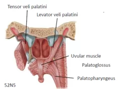
Tensor Veli Palatini Muscle Anatomy
Tensor veli palatini muscle is a broad, thin, ribbon-like muscle in the head that tenses the soft palate.
Along with the levator veli palatini, palatoglossus, palatopharyngeus, musculus uvulae, salpingopharyngeus, stylopharyngeus, superior pharyngeal constrictor, middle pharyngeal constrictor, and inferior pharyngeal constrictor, it is a member of the soft palate and pharyngeal muscle group.
A muscle’s name might reveal a lot about its attachments and purpose. The Latin verb tendere, which means “to stretch,” is where the name tensor originates. The Latin term “veli palatini” means “veil or curtain of the palate.” This etiology clarifies the muscle’s purpose, which is to tighten the soft palate’s palatine aponeurosis and open the auditory tube’s pharyngeal aperture while swallowing or yawning.
Origin
Medial pterygoid plate of the sphenoid bone (scaphoid fossa).
The tendon that runs around the medial side of the pterygoid hamulus of the sphenoid bone is where the muscle fibers merge inferiorly. The tensor veli palatini muscle uses the pterygoid hamulus as a trochlea or pulley. It plays a crucial role in both producing the muscle’s contractile force and horizontally rerouting the pull of the muscle. Tensor veli palatini tendon inserts into the palatine aponeurosis, which makes up the anterior section of the soft palate, after passing the pterygoid hamulus.
Insertion
Palatine aponeurosis.
Dilator tubae component
A portion of the muscle fibers attach to Ostmann’s fat pad, the lateral lamina of the cartilaginous portion of the pharyngotympanic tube, and the surrounding connective tissue.
The dilator tube is the term used to refer to the part of the muscle that has these attachments.
Nerve Supply
Medial pterygoid of mandibular nerve (V3).
It is the sole palate muscle that is not innervated by the vagal and glossopharyngeal nerves, which comprise the pharyngeal plexus.
Blood supply
Both the ascending palatine branch of the facial artery and the larger palatine branch of the maxillary artery provide arterial blood to the tensor veli palatini.
Actions:
Tension of the soft palate.
Function
The anterior part of the soft palate is tensed and depressed by bilateral contraction of the tensor veli palatini. Pulling the soft palate to one side is the result of unilateral muscular contraction. Furthermore, while swallowing or yawning, the tensor veli palatini opens the auditory tube, balancing the pressure (or “popping the eardrums”) between the middle ear and the nasopharynx.
Relations
Tensor veli palatini is located in the pterygoid fossa, between the lateral and medial pterygoid plates, together with the medial pterygoid muscle. The proximal portion of the medial pterygoid muscle is connected to the lateral surface of the tensor veli palatini near its starting point. Tensor veli palatini pierces the buccinator muscle near its attachment point on a tendinous band known as the pterygomandibular raphe en route to the soft palate.
There are numerous significant connections between the head’s neurovascular tissues and the tensor veli palatini. The mandibular, auriculotemporal, and chorda tympani nerves are connected to the lateral surface of the tensor veli palatini in the infratemporal fossa, along with the middle and accessory meningeal arteries. Furthermore, the otic ganglion and the cartilaginous portion of the auditory canal are divided by the tensor veli palatini.
FAQ
Tensing the soft palate, the tensor veli palatini helps the levator veli palatini raise the palate during swallowing, occludiing and blocking the passage of food into the nasopharynx.
The palatine aponeurosis is where the tensor veli palatini muscle, which originates at the medial pterygoid plate of the sphenoid, attaches. In order to stop food from entering the nasopharynx during swallowing, the tensor veli palatini muscle tenses the soft palate.
The main muscle involved in velar elevation is thought to be the levator veli palatini, or levator. It closes the gap between the nasal and oral chambers by raising the velum, as its name suggests.
The masticatory muscles, mylohyoid, anterior belly of the stomach, tensor veli palatani, and tensor tympani originate from the first arch and are innervated by the trigeminal nerve’s branches.
The mandibular nerve gives rise to the nerve to tensor veli palatini, which is a branch of the nerve to medial pterygoid. The foramen ovale allows the mandibular nerve to descend and enter the infratemporal fossa. Here, the medial pterygoid muscle receives a neuronal branch from the mandibular nerve’s main stem.
References
- Tensor veli palatini muscle. (2023, August 31). In Wikipedia. https://en.wikipedia.org/wiki/Tensor_veli_palatini_muscle
- Sendić, G. (2023, October 30). Tensor veli palatini muscle. Kenhub. https://www.kenhub.com/en/library/anatomy/tensor-veli-palatini-muscle
One Comment