Phrenic Nerve Palsy: Causes, Symptoms, Treatment & Physiotherapy
Table of Contents
What Is Phrenic Nerve Palsy?
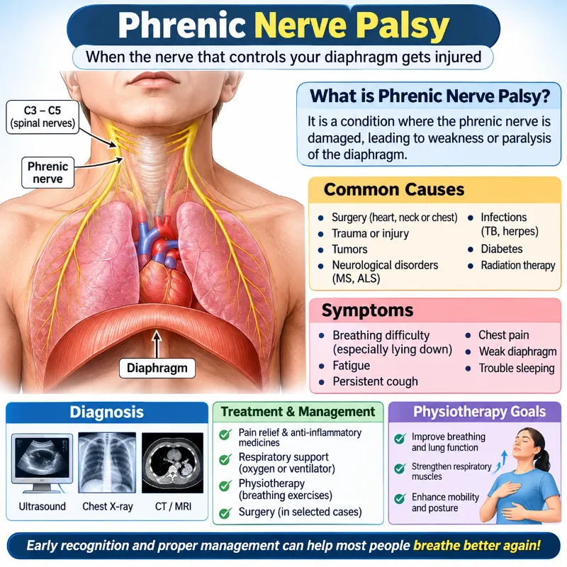
Phrenic nerve palsy is a neurological condition that causes damage or dysfunction to the phrenic nerve. The phrenic nerve is extremely important for respiration due to providing motor innervation to the diaphragm.
If the phrenic nerve is damaged, the diaphragm may become weakened or partially/fully paralysed. This can cause the affected part of the diaphragm to fail to work correctly, causing decreased expansion of the lungs.
Phrenic nerve palsy can occur unilaterally on one side or bilaterally on both sides. Depending on how much damage has occurred to the nerve and how many sides are affected, the symptoms will vary.
The phrenic nerve runs from the cervical area of the spinal cord down the neck and chest to the diaphragm. Damage can occur anywhere along the nerve.
What Is The Phrenic Nerve?
The phrenic nerve is a bilateral mixed peripheral nerve. It is the major nerve controlling the diaphragm.
It originates mainly from the anterior (ventral) rami of the 3rd, 4th, and 5th cervical nerves (C3-C5). Most commonly, the 4th cervical nerve provides the majority.
Anatomists often remember how it originates by using the phrase:
C3, C4 and C5 keep the diaphragm alive.
Origin of the Phrenic Nerve
- C3 – Third cervical spinal nerve.
- C4 – Fourth cervical spinal nerve.
- C5 – Fifth cervical spinal nerve.
Nerve Supply of the Phrenic Nerve
The phrenic nerve provides the following innervation:
1. Motor supply
- Supplies the diaphragm on the same side.
- Controls diaphragmatic contraction during inspiration.
2. Sensory supply
It carries sensory information from:
- The central portion of the diaphragmatic pleura.
- The mediastinal pleura.
- The fibrous pericardium.
- The parietal layer of the serous pericardium.
- The central portion of the diaphragmatic peritoneum.
Important anatomical point: The peripheral portion of the diaphragm receives sensory innervation mainly from the lower intercostal nerves, not the phrenic nerve.
Types and Location of the Phrenic Nerve
There are two phrenic nerves:
1. Right phrenic nerve
- Originates from C3–C5.
- Descends through the neck, passing over the anterior surface of the anterior scalene muscle.
- Enters the thorax and travels along the right side of the heart and pericardium.
- Passes through the diaphragm near the inferior vena cava.
- Supplies the right half of the diaphragm.
2. Left phrenic nerve
- Also originates from C3–C5.
- Descends along the anterior surface of the left anterior scalene muscle.
- Enters the thorax and travels over the left side of the pericardium.
- Passes through the muscular portion of the diaphragm.
- Supplies the left half of the diaphragm.
Functions of Phrenic Nerve
The phrenic nerve is essential for coordinating the diaphragm’s movements during breathing. The main function is controlling the movement of the diaphragm.
Its main functions are:
- Diaphragmatic contraction: It activates the diaphragm during inspiration, allowing the muscle to contract and move downward.
- Lung expansion: Diaphragmatic contraction increases the volume of the thoracic cavity, helping air enter the lungs.
- Sensory function: It carries sensory information from the central diaphragm, pericardium, and adjacent pleural and peritoneal structures.
- Respiratory support: It helps maintain effective breathing, particularly during quiet respiration.
During inspiration, the diaphragm will contract and descend, increasing the vertical size of the thoracic cavity. When the diaphragm relaxes, it will ascend, assisting in expiration.
Causes of Phrenic Nerve Palsy
Phrenic nerve palsy can occur following injury, compression, inflammation, or neurological disease of the nerve. Some patients may find the cause of their phrenic nerve palsy, whilst others may have idiopathic causes.
Common causes include:
Surgical injury
Sometimes the phrenic nerve can get injured or stretched during Thoracic, cardiac, or cervical surgery. There is a higher risk during surgery involving the heart, lungs, mediastinum or neck.
Trauma
Blunt or penetrating injuries to the neck, chest or cervical spine can cause injury to the phrenic nerve. This can occur following road traffic accidents or other serious injuries.
Tumours/Cancer
Tumours in the neck, lung apex or mediastinum may cause compression or damage to the nerve. Some cancers can spread to surrounding areas, causing damage to the nerve.
Neurological Disorders
Disorders involving the cervical spine, nerve roots or peripheral nerves can cause damage to the phrenic nerve. This can include: multiple sclerosis, amyotrophic lateral sclerosis (ALS), cervical spinal cord disorders, cervical nerve root lesions.
Infection
Some infections can cause damage or inflammation to the phrenic nerve. This could include herpes zoster or Lyme disease.
Metabolic disorders
Damage to peripheral nerves can occur in diabetes mellitus, contributing to phrenic nerve neuropathy. However, this is uncommon as the phrenic nerve is usually not isolated.
Autoimmune/Inflammatory disorders
Immune-mediated neurological disorders such as Guillain-Barré syndrome or neuralgic amyotrophy (Parsonage-Turner syndrome) can affect the phrenic nerve.
Radiotherapy
Nerve damage can occur following radiotherapy to the neck or chest. This can happen months or years after treatment.
Cervical Spine Disorders
There can be disorders in the cervical spine that can affect the nerve signals going to the diaphragm. This can include degenerative diseases or injury to the C3-C5 nerve roots.
Idiopathic
In some patients, no cause is found. This is deemed idiopathic.
Epidemiology
Phrenic nerve palsy is a rare condition, and the true prevalence in the general population is unknown.
It can occur following cardiac or thoracic surgery. or after traumatic injury, neurological disorders, or certain infections. It can occur at any age and in both sexes.
Injury to the diaphragm can be found in patients with severe blunt or penetrating trauma. However, this is not the same as diaphragmatic rupture and phrenic nerve palsy.
Risk Factors
There are several risk factors for phrenic nerve dysfunction.
- Trauma – serious injury to the neck, chest or cervical spine.
- Previous Surgery – cardiac, thoracic or cervical surgery.
- Neurological Disorders – ALS, MS, peripheral neuropathy.
- Infection – Some viral or bacterial infections of the nervous system.
- Tumour – Tumours in the cervical area, lung apex or mediastinum.
- Radiotherapy – Previous radiotherapy to the neck/chest.
- Autoimmune Disorders – Conditions causing inflammation/damage to peripheral nerves.
- Diabetes Mellitus – If a patient has had diabetes for a long period of time, this can cause damage to nerves.
- Unknown – Some patients will have no identifiable risk factor.
Symptoms
The symptoms of phrenic nerve palsy depend on whether it is unilateral or bilateral and how much diaphragmatic dysfunction there is.
Unilateral phrenic nerve palsy can be asymptomatic in some patients. Others may have difficulty breathing with physical exertion.
Common symptoms include:
- Shortness of breath – Difficulty breathing especially with exertion, exercise or climbing stairs.
- Orthopnoea – Breathlessness which worsens when lying flat and improves when sitting or standing up.
- Fatigue – Poor oxygenation and increased effort to breathe can cause fatigue.
- Sleep disturbance – Difficulty breathing when sleeping can cause frequent waking, poor quality of sleep or drowsiness.
- Chronic cough – Some patients may find that they have a weak cough due to reduced respiratory muscle function.
- Poor exercise tolerance – Physical activities can be difficult as respiratory muscles are unable to provide sufficient ventilation.
- Chest pain – Some patients experience chest pain.
- Paradoxical abdominal movement – During inspiration, the abdomen can go in rather than out. This is more common with bilateral diaphragm weakness.
- Respiratory infections – Reduced lung expansion and ineffective cough can increase risk of respiratory complications in some patients.
Unilateral Phrenic Nerve Palsy
If there is only one phrenic nerve affected, the other side may still be functioning. Many patients will present with mild symptoms or be asymptomatic.
Symptoms may include:
- Shortness of breath on exertion.
- Poor exercise tolerance.
- Mild orthopnoea.
- Raised diaphragm on X-ray of the affected side.
Bilateral Phrenic Nerve Palsy
If both nerves are affected, movement of the diaphragm can be severely affected.
Symptoms may include:
- Severe shortness of breath even with minimal exertion.
- Difficulty breathing when lying flat.
- Breathing difficulties when sleeping.
- Headaches on waking due to carbon dioxide retention at night.
- Weak cough.
- Low oxygen levels or high carbon dioxide levels in severe cases.
- Respiratory failure in severe cases.
Generally, symptoms will be more severe due to both sides being affected.
Diagnosis
To diagnose phrenic nerve palsy, a doctor will assess the symptoms, medical history, physical examination, respiratory function, and diaphragm function.
Several tests may be performed to determine if the diaphragm is weakened or paralysed and to determine the cause of the condition.
Physical Examination
A healthcare professional will look at your breathing pattern, chest expansion, and abdominal movement.
They may look at:
- Chest and abdominal movement during inspiration and expiration.
- Respiratory rate and breathing effort.
- Oxygen levels via pulse oximetry.
- Breathing sounds using a stethoscope.
- Breathing difficulties in various positions.
- Signs of respiratory muscle weakness.
If one side of the chest is not moving as well or there is unusual abdominal movement, it may be suspected that there is dysfunction of the diaphragm.
Chest X-ray (Chest Radiography)
A chest X-ray is commonly used as an initial investigation when diaphragmatic paralysis is suspected.
It may demonstrate:
- Elevation of one hemidiaphragm.
- Abnormal positioning of the diaphragm.
- Reduced lung volume on the affected side.
- Associated lung abnormalities.
Raised hemidiaphragm can also occur secondary to other conditions including reduced lung volumes, abdominal masses and diaphragm eventration.
- Ultrasound
Ultrasound is an invaluable non-invasive tool.
It is used to visualise the diaphragm moving during respiration and to measure the thickness of the diaphragm.
It can help identify reduced diaphragm excursion, no diaphragm movement, paradoxical movement during inspiration, reduced thickness, and reduced thickening during inspiration. Ultrasound can also be used to monitor the progression of the diaphragm over time.
- Fluoroscopy (Sniff Test)
In this test, patients are asked to make a short, forceful inhale through their nose whilst the diaphragm is viewed during real-time X-ray.
During normal respiration, the diaphragm will descend.
With diaphragm paralysis, the affected side may rise during inspiration. This is known as paradoxical motion.
This is especially useful to identify unilateral diaphragm paralysis.
- Pulmonary Function Tests (PFTs)
PFTs can help to identify how much the diaphragm weakness is affecting respiration.
Common issues seen include reduced forced vital capacity (FVC), reduced lung volumes, restrictive pattern, and reduced FVC when changing from sitting to supine. PFTs can be useful to help identify diaphragm weakness by comparing results between sitting and supine.
CT scans can provide detailed images of the chest and surrounding structures.
It can help identify the cause of the phrenic nerve palsy. This may include lung tumours, mediastinal masses, enlarged lymph nodes, structural problems, lung disease and diaphragm abnormalities. It can also help identify if there is any muscle atrophy or thinning. Ultrasound and MRI can provide more information.
- MRI
MRI can be recommended if a lesion is suspected in the cervical spine, nerve roots, neck or mediastinum.
It can help identify cervical spine abnormalities, nerve root compression, soft tissue tumours, brachial plexus abnormalities and lesions affecting the phrenic nerve pathway. This is especially useful if detailed soft tissue imaging is required.
- Phrenic Nerve Conduction Studies/Electromyography (EMG)
These are used to help identify the electrical activity within the nerve and diaphragm.
They can help to demonstrate nerve conduction issues, identify the electrical activity within the diaphragm, differentiate between nerve damage and muscle disease, and identify the extent of the neurological issues. They are usually carried out by specialists and are used to provide extra confirmation.
Differential Diagnosis
There are several causes that can lead to a raised diaphragm or similar symptoms to phrenic nerve palsy. It is therefore important to identify what the underlying issue is.
Common differentials include:
Diaphragm Eventration
This can occur when part or all of the diaphragm becomes abnormally raised due to thinning or abnormal development of the muscle. It is not necessarily caused by injury to the nerve, like acquired paralysis.
Reduced Lung Volumes
Conditions causing reduced lung volumes can cause the diaphragm to rise. These can include:
- Lung collapse (atelectasis)
- Pulmonary Fibrosis
- Previous lung surgery, e.g., lobectomy
- Radiation-induced lung fibrosis
Abdominal Masses
Masses within the abdomen can cause the diaphragm to rise. These can include:
- Liver enlargement or liver masses
- Splenomegaly
- Abdominal tumours
- Subphrenic abscess/Fluid collection
Increased Abdominal Pressure
Excess fat within the abdomen, ascites, or other conditions causing increased intra-abdominal pressure can cause the diaphragm to rise.
Diaphragm Injury
Trauma or structural issues can cause issues with the normal function of the diaphragm.
Pleural/Pulmonary Disease
Some pleural or pulmonary diseases can cause the diaphragm to be abnormally positioned or cause reduced respiration.
Treatment
Treatment will depend on the underlying cause, severity of the diaphragm weakness, respiratory symptoms, and if it is affecting one or both sides.
The aim is to improve respiration, control the underlying cause, prevent respiratory complications, and try to regain full function of the diaphragm.
Medical Treatment
Treatment will be focused around the underlying cause. If the patient is experiencing pain due to nerve damage or associated conditions, appropriate analgesia may be prescribed. If the underlying cause is due to infection, then antibiotics may be required.
If it is due to an inflammatory condition, then specific anti-inflammatory medications may be considered for select autoimmune conditions. Underlying conditions such as diabetes and respiratory disease should be well controlled.
Corticosteroids, muscle relaxants, or other medications are not routinely used to treat phrenic nerve palsy, however depending on the underlying cause.
Respiratory Support
Patients with significant diaphragm weakness may require additional respiratory support.
Non-Invasive Ventilation (NIV)
NIV can be used to assist respiration via a mask. It may be recommended for patients with nocturnal hypoventilation, CO2 retention, or respiratory insufficiency.
Oxygen Therapy
Supplementary oxygen may be required if the patient has low oxygen levels in their blood. Oxygen will not help with poor ventilation or CO2 retention.
Ventilation
For severe cases, invasive ventilation may be required.
Surgical Treatment
Surgery may be considered if symptoms are still significant following conservative treatment or if there is a specific surgical fix for a nerve injury.
The main surgical options include:
Diaphragmatic plication
Diaphragmatic plication is a surgical procedure indicated for selected patients with symptomatic unilateral diaphragmatic paralysis.
The raised and weak diaphragm is folded and sutured.
This helps reduce paradoxical breathing, increase the efficiency of lung expansion, improve respiratory efficiency, reduce breathlessness, and improve exercise tolerance. This treatment does not directly treat the damaged phrenic nerve, but improves the mechanics of the diaphragm.
B. Phrenic nerve repair
Surgical repair may be considered in selected patients with an identifiable phrenic nerve injury.
Depending on the type and extent of the nerve injury, the surgeon may choose to undertake nerve decompression, nerve repair, nerve graft or nerve transfer. A nerve graft may be used if the damaged section of nerve cannot be directly repaired. The sural nerve can be used as this is a sensory nerve located in the lower limb.
The aim of this treatment is to improve nerve continuity and encourage the return of diaphragmatic muscle activity. This occurs gradually and may take many months.
C. Diaphragmatic pacing
Electrical stimulation of the phrenic nerve/diaphragm.
This may be considered for very carefully selected patients who have certain causes of ventilatory failure. The nerve and diaphragm must be able to respond to stimulation.
This treatment is not suitable for everyone with a phrenic nerve palsy. A thorough assessment of nerve conduction and diaphragmatic muscle function must be carried out.
Potential Complications of Surgical Treatment
Like any surgical treatment, there can be complications. These can include:
- Bleeding
- Surgical site infection
- Damage to adjacent nerves and vessels
- Persistent respiratory problems
- Pneumonia
- Discomfort/pain
- Failure to improve
- Symptoms returning
The risk of complications depends on the type of surgery, the patient’s general health, and the extent of their underlying problem.
Physiotherapy Management of Phrenic Nerve Palsy
Physiotherapy is an important treatment modality within the management of patients with a phrenic nerve palsy. Patients will benefit from respiratory rehabilitation to improve breathing efficiency, increase chest wall mobility, increase their exercise tolerance and reduce complications.
The treatment should be tailored to the patient depending on the severity of nerve damage, respiratory function, exercise tolerance and overall general health.
Aims
- To improve the patient’s respiratory function.
- Improve lung expansion.
- Improve chest wall mobility.
- Reduce respiratory fatigue.
- Increase exercise tolerance.
- Maintain upper body strength and mobility.
- Reduce complications from impaired ventilation.
- Improve posture and physical activity.
- Encourage independence.
A. Breathing Exercise
Breathing exercises are an important part of respiratory rehabilitation. They can help improve control and increase the efficiency of the respiratory muscles the patient has.
Diaphragmatic breathing
Lie comfortably on your back with knees slightly bent. Place 1 hand on your upper chest and 1 hand on your abdomen. Inhale slowly through your nose. Let the abdomen rise whilst keeping the chest quite still. Exhale slowly through your mouth. Repeat gently.
- Improves patient awareness of their breathing.
- Encourages efficient use of the available diaphragmatic function.
- Helps to reduce accessory muscle use.
- Encourages relaxation.
Note: Diaphragmatic breathing will not help to improve nerve function in a fully paralysed diaphragm. The exercise should be adapted to the amount of muscle activity the patient actually has.
Sit comfortably with relaxed shoulders. Inhale slowly through your nose. Pucker your lips as if on a gentle out. Exhale slowly through pursed lips. Do not force air out of your lungs. Repeat several times.
- Helps to control breathing.
- Encourages controlled exhale.
- May help reduce feelings of breathlessness.
Incentive Spirometry
An incentive spirometer is used to encourage slow deep breaths.
Sit up in a comfortable position. Hold the incentive spirometer upright. Exhale normally. Place your lips firmly around the mouthpiece. Inhale slowly and deeply. If comfortable, hold your breath for a short time. Remove the mouthpiece and exhale normally. Repeat as instructed.
Encourages deep breathing.
Helps to keep lungs expanded.
May help reduce post-operative pulmonary complications. This does not directly improve nerve function or strengthen the diaphragm.
B. Chest Expansion Exercises
Chest expansion exercises encourage ribcage movement.
Sit upright with back supported. Place your hands around your lower ribs. Inhale slowly through your nose. Feel your ribcage expand. Exhale slowly. Repeat as tolerated.
- Encourages chest wall movement.
- Helps to maintain thorax mobility.
- Encourages controlled breathing.
- May improve awareness of respiratory movements.
C. Thoracic Mobilisation Exercises
Due to reduced activity and altered breathing patterns, the thoracic spine and chest wall may become stiff.
Thoracic mobilisation can help maintain normal upper body movement.
Thoracic extension
Sit in a comfortable position on a chair. Place your hands behind your head. Gently arch your upper back over the chair if comfortable. Keep your neck relaxed. Return to start position. Repeat gently.
Shoulder Mobility Exercises
Sit up with your feet on the ground. Cross your arms across your chest. Gently rotate your upper body to one side. Return to centre. Repeat on the other side.
Benefits:
- Helps maintain spinal mobility.
- Improves upper body mobility.
- Reduces stiffness.
- Encourages comfortable chest wall movement.
D. Shoulder Mobility Exercises
Shoulder mobility exercises can help maintain upper body movement and reduce stiffness due to reduced activity.
- Shoulder rolls
- Shoulder flexion
- Shoulder abduction
- Wall climbing
- Gentle arm circles
These exercises help maintain shoulder mobility. This does not improve nerve function.
E. Respiratory Muscle Training
Inspiratory muscle training can be considered if appropriate.
This uses a special device to provide resistance on inhalation.
This can help improve inspiratory muscle strength and stamina, improve breathing efficiency, and improve exercise tolerance in selected patients. The resistance should be decided by a physiotherapist or respiratory specialist following an assessment of respiratory muscle function.
Patients with severe diaphragmatic paralysis should not commence resistance breathing exercises without professional assessment.
F. Upper Body and General Strengthening Exercises
General strengthening exercises can help maintain muscle strength.
- Shoulder flexion
- Shoulder abduction
- Elbow flexion/extension
- Resistance band exercises
- Light functional strengthening
Start with a comfortable intensity and build up gradually depending on respiratory capacity.
G. Aerobic Exercise
Aerobic exercise can help improve cardiovascular fitness and exercise tolerance.
- Walking
- Stationary cycling
- Gentle low-impact exercises
- Supervised endurance exercises.
Intensity and duration should be adjusted depending on oxygen saturation, breathlessness, heart rate and overall exercise tolerance.
Patients with significant respiratory problems may require supervised pulmonary rehab.
H. Postural Advice
- Correct posture can help reduce breathing difficulties.
- Sit up when experiencing breathlessness.
- Use pillows to prop up your upper body when sleeping.
- Avoid lying flat for long periods of time.
- Sit and stand with a comfortable posture.
- Take regular breaks to move.
Some patients with diaphragmatic weakness find it easier to breathe when in a semi-reclining or upright position.
Precautions
Physiotherapy should be undertaken carefully as patients may have reduced respiratory capacity.
- Check how difficult the patient is breathing.
- Oxygen saturation should be monitored if indicated.
- Avoid over-exertion.
- Take adequate breaks between exercises.
- Avoid holding breath or straining.
- If the patient experiences severe breathlessness, dizziness, chest pain or unusual tiredness, stop the exercise.
- Follow medical advice on supplementary oxygen and ventilatory support.
- Avoid any exercises causing significant respiratory distress.
Patients with bilateral diaphragmatic paralysis or respiratory failure should have a specialist respiratory assessment before undertaking any exercise.
Complications of Phrenic Nerve Palsy
If diaphragmatic dysfunction is severe or left untreated, there can be several respiratory complications.
Common complications include:
- Respiratory insufficiency – Reduced diaphragmatic activity can cause ventilation issues and affect oxygen and carbon dioxide levels.
- Hypoventilation at night – Reduced respiratory muscle activity can cause hypoventilation whilst sleeping.
- Atelectasis – Reduced lung expansion can cause partial collapse of the lungs.
- Frequent respiratory infections – Poor coughing and secretions can increase the risk of respiratory infections.
- Exercise intolerance – Reduced respiratory capacity can cause reduced exercise tolerance.
- Chronic respiratory failure – Severe bilateral diaphragmatic dysfunction can cause long-term ventilation issues.
- Reduced quality of life – Breathlessness and disturbed sleep can affect everyday activities.
Prognosis of Phrenic Nerve Palsy
The prognosis depends on the cause of nerve injury, severity of diaphragmatic dysfunction, number of affected sides, and the patient’s general health.
Patients with unilateral phrenic nerve palsy often have a relatively good prognosis. If the other side of the diaphragm is functioning normally and there are no significant underlying lung or heart problems.
Some patients will gradually regain diaphragmatic function. This can happen if the nerve injury was temporary or due to a reversible issue.
However, if there was severe nerve damage, there may not be a full recovery.
Bilateral palsy is generally a more serious prognosis as both sides of the diaphragm are affected. Patients may require long-term respiratory support and regular monitoring.
Early diagnosis, correct management of the underlying cause, and a respiratory rehabilitation programme can help improve functional outcomes and quality of life.
When to seek medical attention
Seek immediate medical attention if you are having:
- Sudden or severe breathing difficulties.
- Blue or grey lips/fingertips.
- Confusion or unusual drowsiness.
- Severe chest pain.
- Worsening of breathing difficulties.
If you are experiencing ongoing breathlessness, difficulty lying flat, unusual disturbances to sleep or reduced exercise tolerance, then you should seek medical attention.
Conclusion
Phrenic nerve palsy is a neurological condition affecting the nerve that controls the diaphragm. Damage to this nerve can reduce diaphragmatic activity and impact normal breathing.
Early diagnosis can help determine the cause and reduce risk of complications. Treatment can include medical management, respiratory support, surgery and physiotherapy depending on the patient’s condition.
Physiotherapy can help improve breathing efficiency, maintain chest wall mobility, improve exercise tolerance and keep patients active. A tailored rehabilitation programme alongside correct medical management can help improve respiratory function and quality of life.
FAQs
A paralyzed or weakened diaphragm can occur from phrenic nerve palsy, also referred to as phrenic nerve injury, which can cause a variety of symptoms that mostly impact breathing and occasionally cause other difficulties. Depending on whether the paralysis is unilateral (affecting only one side) or bilateral (affecting both sides), these symptoms might vary in severity.
The most common and significant symptom is dyspnea.
Phrenic nerve injury is characterized by breathing difficulties or difficulty breathing, particularly when straining yourself or when lying down.
Breathlessness: Even when at rest or only when exercising, patients may suffer from chronic dyspnea.
Weariness: Chronic fatigue may result from a decreased capacity for effective breathing.
Sleep disturbances: Sleep apnea and other respiratory issues connected to sleep can be brought on by injury to the phrenic nerve.
Shoulder pain: Phrenic nerve irritation may be the cause of unexplained shoulder pain, particularly when accompanied by arm weakness.
Primary lung tumors, malignant mediastinal tumors, and mediastinum surgery are among the many possible causes. It might be idiopathic.
An imaging examination called a sniff test assesses the function of your diaphragm.
An imaging examination called a sniff test assesses the function of your diaphragm. As you breathe in and out, a healthcare professional captures your chest X-ray pictures. The pictures may indicate if you have paralysis or weakening in your diaphragm.
It can be done for reconstructive plastic surgeons to reverse diaphragm paralysis because of advances in nerve decompression and transplantation. Surgeons have been able to restore function to previously paralyzed muscle groups by using techniques that are developed from typical procedures used to treat arm or leg paralysis.
Its movements are controlled by the phrenic nerve, which gives the diaphragm signals from the brain to help with breathing control. If something stressful produces sudden excitement, anxiety, fear, or shock, the diaphragm may begin to spasm.
Thoracic or otolaryngology surgeons will come into contact with the phrenic nerve, but they will need to avoid it because it is not frequently handled outside of neck or chest surgery.
In addition, there has been increasing evidence that SARS-CoV-19 may infiltrate peripheral nerve terminals. It implies that COVID-19 patients might suffer phrenic paralysis as a result of the virus, which could lead to pulmonary decompensation.
Phrenic nerve palsy is caused by supraclavicular brachial plexus block in 67% of situations. It is probable that a nearby local anesthetic temporarily disrupts the nerve’s normal function.
Reconstructive plastic surgeons can now reverse diaphragm paralysis due to developments in nerve decompression and transplantation. Surgeons have been able to restore function to previously paralyzed muscle groups by using techniques that are developed from typical procedures used to treat arm or leg paralysis.
Its movements are controlled by the phrenic nerve, which gives the diaphragm signals from the brain to help with breathing control. The diaphragm may begin to spasm if a stressful situation causes abrupt excitement, anxiety, dread, or shock.
My areas of expertise are head and neck surgery and plastic surgery. Thoracic or otolaryngology surgeons will come into contact with the phrenic nerve, but they will need to avoid it because it is not frequently handled outside of neck or chest surgery.
In addition, there has been increasing evidence that SARS-CoV-19 may infiltrate peripheral nerve terminals. It implies that COVID-19 patients might suffer phrenic paralysis as a result of the virus, which could lead to pulmonary decompensation.
Phrenic nerve palsy is a common result of supraclavicular brachial plexus block in 67% of cases. It is probable that a nearby local anesthetic temporarily disrupts the nerve’s normal function. As you breathe in and out, a healthcare professional captures your chest X-ray pictures.
References:
- Mandora, S., & Mead, T. (2023, August 20). Phrenic nerve injury. StatPearls – NCBI Bookshelf. https://www.ncbi.nlm.nih.gov/books/NBK482227/
- https://www.hackensackmeridianhealth.org/en/healthu/2024/02/02/when-phrenic-nerve-damage-causes-diaphragm-muscle-paralysis-how-to-spot-it
- Bell, D., & Gaillard, F. (2008b). Phrenic nerve palsy. Radiopaedia.org. https://doi.org/10.53347/rid-1869
- Sankhla, D. (2023c, August 19). Phrenic nerve palsy – Cause, symptoms, treatment. Samarpan Physiotherapy Clinic. https://samarpanphysioclinic.com/phrenic-nerve-palsy/