
Diaphragm Muscle
Table of Contents
Introduction
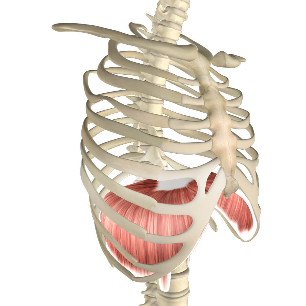
The diaphragm is an unpaired, dome-shaped skeletal muscle that is located inside the trunk. It is the primary muscle that is active in inspiration. Contraction of the diaphragm muscle facilitates the expansion of the thoracic cavity.
This increases the volume of the cavity, which in turn reduced the intrathoracic pressure allowing the lungs to expand and inspiration to occur.
The diaphragm is much more than just a sheath separating your thoracic cavities and abdominal cavities.
Origin of Diaphragm
The sternal part of the diaphragm originates from the posterior aspect of the xiphoid process
The coastal part of the diaphragm originates from the Internal surfaces of lower costal cartilages and ribs 7-12
The lumbar part of the diaphragm originates from the Medial and lateral arcuate ligaments (lumbocostal arches), bodies of vertebrae L1 to L3 (and intervertebral discs), anterior longitudinal ligament
Insertion of Diaphragm
The diaphragm is inserted into the Central tendon.
Nerve supply
The nerve supply of the diaphragm is the Phrenic nerves (C3 to C5)
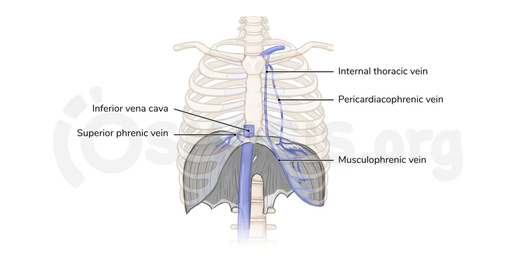
Blood supply
The blood supply of the diaphragm is the Subcostal and lowest 5 intercostal arteries, inferior phrenic arteries, superior phrenic arteries
Action
Depression of the costal cartilages, the primary muscle of the inspiration
Clinical Significance of Diaphragm
Paralysis of the Diaphragm
Diaphragmatic paralysis is because by the interruption in the nerve supply. This may occur in the phrenic nerve, cervical spinal cord, or brainstem. It is most commonly due to a lesion of the phrenic nerve:
Mechanical trauma: ligation or injury to the nerve during surgery.
Compression: due to a tumor
Myopathies: such as myasthenia gravis.
Neuropathies: such diabetic neuropathy.
Paralysis of the diaphragm muscle produces a paradoxical movement. The affected side of the diaphragm muscle moves upwards during inspiration, and downwards during expiration. Unilateral paralysis of the diaphragm is usually asymptomatic and is most often an incidental finding on x-ray. If both sides are paralyzed, the patient may have orthopnoea and fatigue.
Diaphragmatic hernia
A diaphragmatic hernia occurs when an organ in your abdomen bulges into your chest cavity. diaphragmatic hernias can be present at birth or they can result from trauma, age, and obesity. Hernias can require surgical repair.
Spasms
During a diaphragm muscle spasm, the diaphragm doesn’t relax and curves back up during expiration. It contracts, causing a cramp in the abdomen. Strenuous exercise may cause this type of spasm. It usually gets better with rest.
Phrenic nerve damage
Nerve damage can result from trauma, cancer, or autoimmune diseases. It may also happen during surgery including heart bypass surgery and lung transplants. A tumor, aortic aneurysm, or cervical spondylosis can compress or damage the Phrenic nerve. Conditions such as HIV and diseases like Lyme disease can cause the nerve to become inflamed.
Exercise of Diaphragm muscle
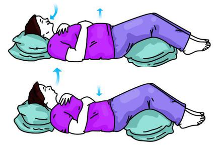
Diaphragmatic breathing exercises
Lie on your back in bed or on a flat surface, with your knees bent and your head supported. You can use a pillow under your both knee to support your legs.
Place one hand on your upper part of the chest and the other just below your rib cage. This will allow you to feel your diaphragm muscle move as you breathe.
slowly Breathe in through your nose so that your stomach moves out, causing your hand to rise. The hand on your upper chest should remain the same as possible.
Tighten your stomach muscles, so that your stomach moves inward, causing your hand to lower as you exhale through pursed lips.
Sandbag Breathing
The patient lies in a supine position. Their spine should be straight. A sandbag is placed on the belly of the patient.
Help the patient to establish a flow of relaxed breathing with the following steps
Feel the breath flowing in and out, repeatedly
Soften the abdomen and feel it rise during inhalation and fall during exhalation
Let the breath flow and don’t pause between breaths
Intercostal Stretching Breath
To perform an intercostal stretching breath, the patient is in a standing position. Ask the patient to stretch both arms over their head. Inhale deeply and on the exhale, they should stretch both arms to the right side. This stretches the intercostal muscles on the left side of the body. As they inhale, they should come back to the starting position.
On the next exhale, they should stretch their arms to the left side, creating a stretch in the right intercostal muscles. Repeat three times on each side.
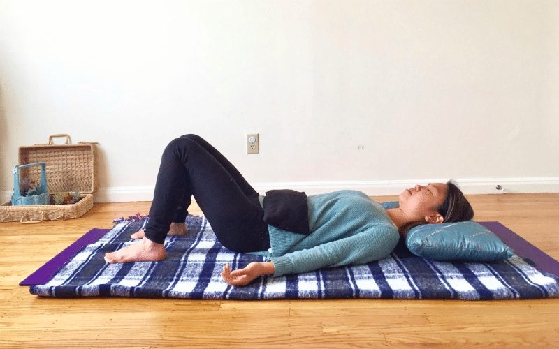
Diaphragm Stretches
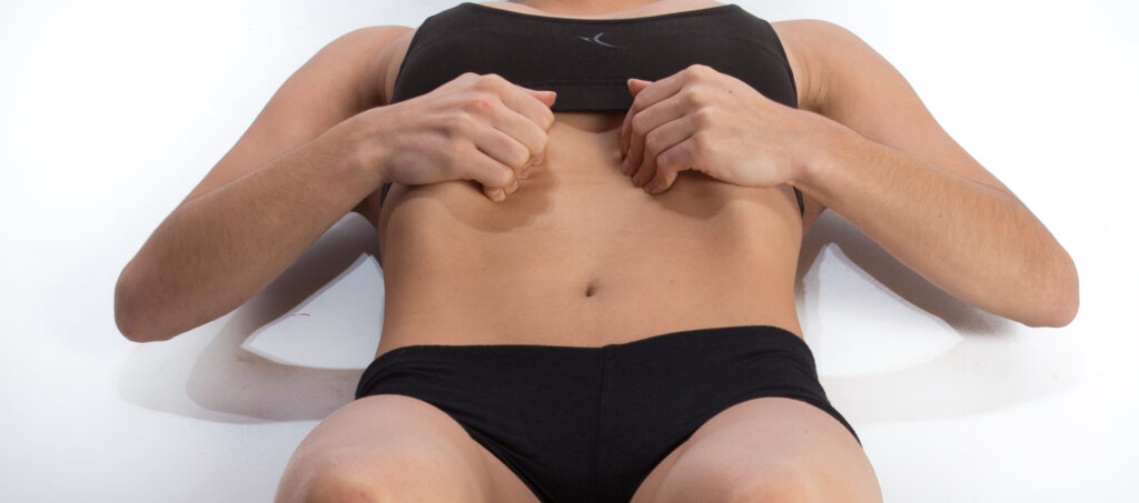
To perform a diaphragm stretch, the patient is in the supine lying position, in 90/90 hip and knee flexion. Place the heels of the patient’s hands on the proximal part of their thighs. They should perform some relaxed breaths through their nose.
After a deep inhalation and full exhalation, the patient should push their hands into their thighs. At this point, they should think about inhaling without actually breathing any air inside. Then ask the patient to suck their belly in and expand their ribs, which creates a vacuum. The patient should then slightly move their spine/pelvis through flexion and extension. lateral shifts will also increase the stretch in different parts of the diaphragm muscle. Breathe normally during one or two cycles and repeat three to four times.
17 Comments