|

Antenatal Exercise and Postnatal exercise

What is Antenatal exercise? ~antenatal exercises are the exercises prescribed during the pregnancy by the physiotherapists.~thsese exercises helps expectant mothers to gain knowledge on how to look after themselves during pregnancy.~antenatal exercises provides many benefits to the pregnant mother if the exercises are performed correctly under the guidance of the physiotherapist and gynaecologists. ~Antenatal exercise…

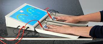
Iontophoresis

Iontophorosis : Indication, Contraindication, Precaution

Introduction:- ~the term iontophoresis is simply defined as ion transfer(ionto=ion),(phoresis=transfer) ~introduction of ions into the body using direct electrical current. ~it is a specialized technique of electrical stimulation that uses electrical polarity of continuous direct current to ionize medicines placed beneath surface electrodes and transfers them into the body through the skin. Basic principle of…

The Cerebrum

Introduction: ~the cerebrum is made of two cerebral hemispheres which are incompletely separated from each other by the median longitudinal fissure.~the two hemispheres are connected to each other across the median plane by the corpus callosum. each hemisphere contains a cavity, called the lateral ventricle. External feautures: three surfaces: 1. the superolateral surface is convex…

The Cerebellum: Anatomy, Function

Introduction: ~The cerebellum is the largest part of the hind brain. it is situated in the posterior cranial fossa behind the pons and medulla.~it is an infratentorial structure that coordinates voluntary movements of the body. Relations: ~Anteriorly:-fourth ventricle,pons and medulla~Posteroinferiorly:-Squamous occipital bone,&~Superiorly:-tentorium cerebelli External features: ~the cerebellum consists of two cerebellar hemisphere that is united…

spinal cord

Spinal Cord

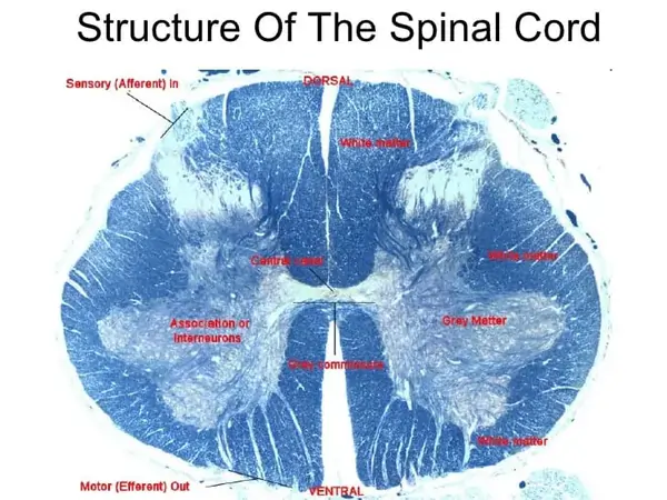
What is the spinal cord? ~the spinal cord is the lower elongated, cylindrical part of the CNS. It occupies the upper two-thirds of the vertebral canal. It extends from the level of the upper border of the atlas to the lower border of vertebra L1 or the upper border of vertebra L2. ~it is about…

Iliococcygeus muscle
| |

Iliococcygeus Muscle

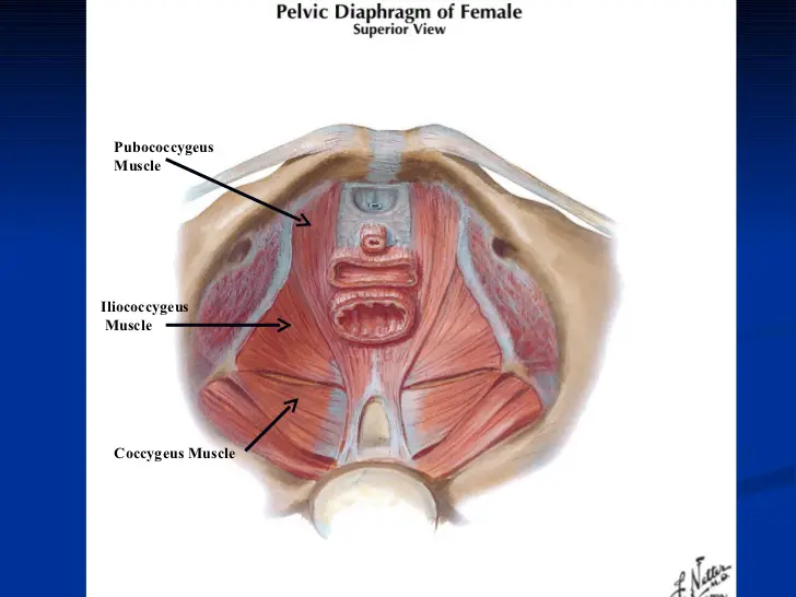
Iliococcygeus Muscle Anatomy The levator ani muscle, which makes up the majority of the pelvic diaphragm, is composed of three muscles, including the iliococcygeus muscle. The flat, broad skeletal muscle is known as the iliococcygeus. the inner side of the ischium (the lower and back part of the hip bone) and from the posterior part…

|

Ankle Joint: Anatomy, Function, Importance

Ankle joints are the most common joints we use in our day-to-day lives, and they’re also the most common joints that require care and attention. Ankle joints are a series of joints that connect your lower leg to your foot, and they’re used for a wide variety of movements and positions. Even the smallest ankle…

Internal oblique muscle
| |

Internal oblique muscle: Origin, Insertion, Function, Exercise

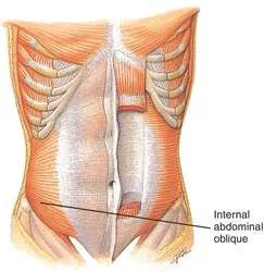
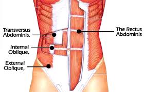
Internal oblique muscle is a thin, broad muscle in the lateral abdominal wall that is located below the external oblique and just above the transverse abdominal muscles. Starting from the superficial layer to the deep layer, the external oblique, internal oblique and transversus abdominis make up the three layers of the lateral abdominal wall. Internal…

External abdominal oblique muscle
| |

External Oblique Muscles

The external oblique is the largest and the most superficial (outermost) of the three flat muscles of the lateral anterior abdomen. Structure: It arises from eight fleshy digitations, each from the external surfaces and inferior borders of the fifth to twelfth ribs. These digitations are arranged in an oblique line that runs inferiorly and anteriorly,…