Biceps Femoris Muscle
Table of Contents
Biceps Femoris Muscle Anatomy
The Biceps Femoris is one of the three muscles that make up the Hamstring group and is used when standing or sitting. The Biceps Femoris is the largest of the three muscles and is responsible for the most movement at the knee. The Biceps Femoris attaches to the ischial tuberosity, lateral side of the head of the fibula, and its main function is to flex the knee.
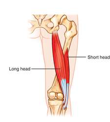
The muscles have two heads, termed as a long head which is located superficially while the ‘short head’ is located deep.
This muscle starts from the ischial tuberosity runs down to the end at the head of the fibula. The muscles cross two joints; the hip joint and the knee joint. That is why muscles work mainly as a Hip extension, External rotation, and knee flexion.
In this article, you will find Muscle related Anatomies such as Origin, Insertion, Function, Exercise, and clinical importance.
A muscle of the thigh located to the posterior, or back. As its name implies, it has two parts, one of which (the long head) forms part of the hamstrings muscle group.
Origin:
- Long head: inferomedial impression of ischial tuberosity, sacrotuberous ligament
- Short head: linea aspera of femur – lateral lip, lateral supracondylar line of femur
Insertion:
The lateral side of the head of the fibula articulates with the back of the lateral tibial condyle.
Nerve supply:
- long head: tibial nerve a part of scitic nerve.
- short head: common fibular nerve also a part of sciatic nerve.
Blood supply:
Inferior gluteal artery, perforating arteries, popliteal artery
Actions:
- Long head function: Knee flexion, Hip extension, laterally rotates lower leg when knee slightly flexed, assists in lateral rotation of the thigh when hip extended
- Short head function: Knee flexion, laterally rotates lower leg when knee slightly flexed
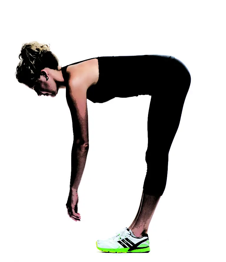
flexes knee joint, laterally rotates knee joint (when the knee is flexed), extends hip joint (long head only).
Relations
The biceps femoris lies superficially in the posterolateral thigh throughout the majority of its length, barely delving deep into the layers of skin, fat, and fascia. The gluteus maximus muscle covers it at its superior aspect, which is the only exception to this rule. The biceps femoris travels across the semimembranosus, adductor magnus, and lateral head of the gastrocnemius muscles as it descends from the pelvis into the posterior thigh area.
It also protects the sciatic nerve by being superficial to it along the route. The common fibular nerve, the terminal branch of the sciatic nerve, is located close to the insertion of the biceps femoris. The nerve follows the tendon as it passes briefly along the medial edge of the biceps femoris. This is a crucial clinical relationship to have when thinking about injuries or doing surgeries in this area.
Clinical Importance
The Biceps femoris two heads are inserted into the head of the fibula, where they divide into two portions by fibular collateral ligaments. Any kind of subluxation or dislocation of this muscle tendon or abnormal insertion of the tendon, trauma, or meniscal instability can lead to Snapping Biceps Femoris Tendon.
It is a rarely occurring condition with symptoms like pain on the lateral side of the knee, over the fibular head will be prominent, a painful snapping sound on the lateral side of the knee during active and passive knee flexion at 90 degrees, and difficulty in performing day-to-day activity.
During sports sprinting there will be a sudden stop-go movement or alter in the speed of running due to which biceps femoris muscle and semitendinosis located at the back of your thigh are usually pulled causing over-stretch of the muscles. The injury to the Long head of the biceps femoris muscle or tendon and semitendinosus are most commonly seen in football and hockey players leading to sport-related injuries.




10 Comments