Temporalis Muscle Anatomy
| | |

Temporalis Muscle Anatomy

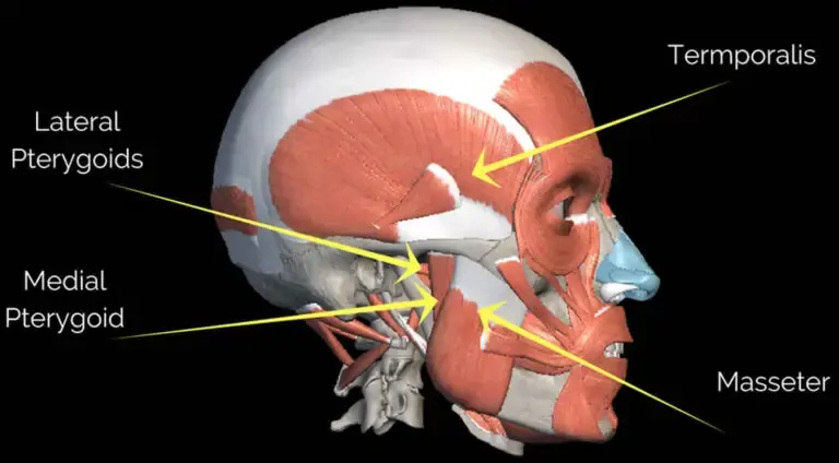
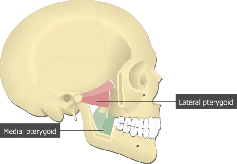
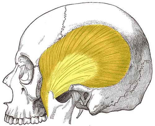
Introduction of the Temporalis muscle In anatomy, the temporalis muscle is also known as the temporal muscle. This muscle fills the temporal fossa. The temporalis muscle is a thin, fan-shaped muscle located within the temporal fossa of the skull. Along with the medial pterygoid muscle, lateral pterygoid, and masseter muscles, it belongs to the group…

The masseter muscle Anatomy
| | |

The masseter muscle Anatomy

Introduction The masseter is a quadrilateral muscle that covers the lateral surface of the ramus of the mandible. The masseter muscle fibres are arranged in three layers. The masseter muscle is a paired, strong, thick, and rectangular muscle that is originating from the zygomatic arch and extends down to the mandibular angle. It contains a…

Medial pterygoid muscle Anatomy
| | |

Medial pterygoid muscle Anatomy

Introduction The medial pterygoid is a thick, quadrilateral muscle of the face and it has a small superficial head and a large deep head which forms the major part of the muscle.The medial pterygoid muscle is supplied by the mandibular branch of the trigeminal nerve (V). It is important in mastication (chewing). It belongs to…

Semispinalis Cervicis
| |

Semispinalis Cervicis

Introduction Semispinalis Cervicis belongs to the semispinalis group of the muscle, which in turn is part of the Transversospinal group of muscles (It is formed of muscles between a spinous process and the vertebrates transverse process below). The semispinalis muscles have the longest fascicles of the transversospinalis group of the muscles, spanning six segments. The…

| |

Middle pharyngeal muscle :

MUSCLE DETAILS: The middle pharyngeal constrictor is a fan-shaped sheet-like muscle of the pharynx. This muscle, along with the superior and inferior pharyngeal constrictors, forms the bulk of the pharyngeal wall. The pharyngeal constrictor muscles create a peristaltic wave that facilitates passage of the food bolus inferiorly, from the pharynx towards the esophagus, during the…

Levatores Costarum Muscles: Anatomy, Origin, Insertion, Function
| | |

Levatores Costarum Muscles: Anatomy, Origin, Insertion, Function

Levatores Costarum Muscles Anatomy Levatores costarum muscles is a twelve in number on either side, are small tendinous and fleshy bundles, which arise from the ends of the transverse processes of the seventh cervical and upper eleven thoracic vertebrae. It is located on either side of the posterior aspect of the thoracic vertebra they go…

Obliquus capitis inferior muscle
| |

Obliquus capitis inferior muscle

Obliquus capitis inferior muscle Anatomy One of the four suboccipital muscles is the obliquus capitis inferior muscle. The largest muscle of the four is obliquus capitis inferior, which is grouped with the rectus capitis posterior major, rectus capitis posterior minor, and obliquus capitis superior muscles. By supporting the atlantoaxial joint, obliquus capitis inferior helps to…

Rectus capitis posterior minor muscle
| |

Rectus capitis posterior minor muscle

Rectus capitis posterior minor muscle Anatomy arises by a narrow pointed tendon from the tubercle on the posterior arch of the atlas, and, widening as it ascends, is inserted into the medial part of the inferior nuchal line of the occipital bone and the surface between it and the foramen magnum, and also takes some…